
A subepithelial connective tissue graft is soft tissue taken from beneath the surface lining of the palate and tucked under the gum to cover an exposed tooth root. For an adult who's noticed a tooth looking longer and felt a sharp ache with cold water, that definition raises two practical questions. What does the graft add, and what does recovery at the palate involve?
Table of Contents
- What a Subepithelial Connective Tissue Graft Is
- When Gum Recession Calls for a Connective Tissue Graft
- How the Graft Is Placed Step by Step
- Recovery at the Palate and Gum Site
- Realistic Results From Published Studies
- How SCTG Compares With Other Gum Graft Options
- Confirm Whether a Graft Fits Your Gums
What a Subepithelial Connective Tissue Graft Is
A subepithelial connective tissue graft, often shortened to SCTG, is a periodontal procedure that places a thin layer of connective tissue over an exposed root and covers it with a repositioned gum flap. It's used to treat gingival recession, the condition in which gum tissue has pulled back and left part of the root uncovered.
Say a lower front tooth looks longer than its neighbors. The gumline sits lower, brushing feels tender at one spot, and cold drinks cause a brief sting. That's the familiar picture of recession, and it's the problem this graft was designed to address.
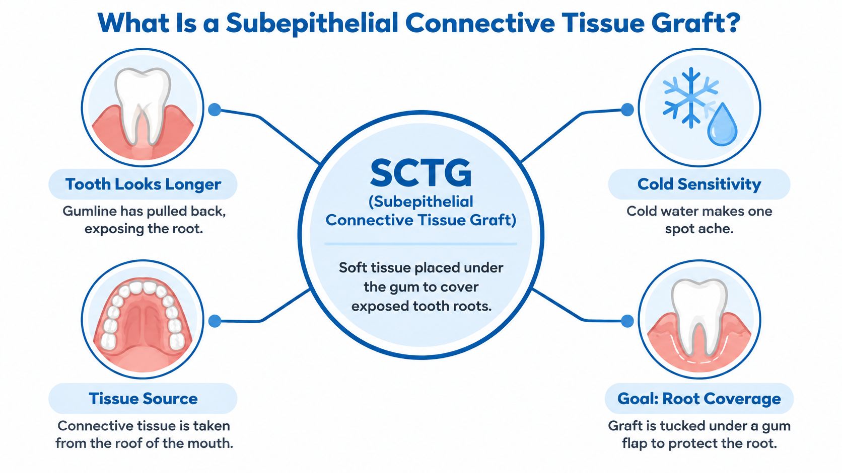
A Simple Way to Picture the Parts
The gum flap works like a blanket. The graft is the padding placed beneath that blanket, adding bulk where the gum is thin or missing so the blanket can cover the bare root more securely. Without padding, a thin blanket may lie flat but offer little protection.
The graft also benefits from what's called a double blood supply. It can draw nourishment from the connective tissue bed beneath it and from the flap above it. That two sided support helps explain why the technique works when the graft is held still and covered well.
The technique was formally described in 1985, when Langer and Langer reported on 56 treated cases followed for up to 4 years, with root coverage ranging from 2 to 6 mm using a partial thickness flap over adjacent recession defects. Those early results belong to that specific group of cases, and individual outcomes vary with the size and position of each defect.
What the Graft Can Do
- Cover part of the exposed root, protecting a surface that wasn't meant to sit in the mouth uncovered.
- Add soft tissue thickness, making thin gum less prone to further breakdown.
- Increase keratinized tissue, the firmer band of gum that tolerates brushing and chewing better.
- Reduce sensitivity that's linked to root exposure, for many patients.
It's often paired with a coronally advanced flap, where existing gum is gently moved toward the crown and secured over the graft. Thickness, protection, and comfort are the primary goals of that combination. Improved appearance tends to follow.
The name describes where the graft comes from: subepithelial means the tissue is taken from beneath the surface layer.
Four labels are worth keeping in mind. The donor site is the palate, the recipient site is the receded gum, the graft is the padding, and the flap is the covering. Choosing this graft means weighing what the tissue adds at the root against a second wound on the palate, and both sides of that trade-off deserve attention.
When Gum Recession Calls for a Connective Tissue Graft
Gum recession is a signal, not a single diagnosis. An exposed root can result from thin tissue, hard brushing, inflammation, tooth position, or a combination of factors. Because the causes differ, two patients with the same amount of visible root can need very different treatment.
Start with the outcome that matters most for this mouth: root coverage, thicker tissue, more keratinized tissue, better appearance, or avoiding a second wound on the palate. That last point deserves honest weight. Every connective tissue graft creates a donor site on the roof of the mouth, so choosing SCTG means accepting a second healing area in exchange for the tissue it delivers. For some mouths that trade is clearly worth it. For others, an alternative material avoids the palate entirely.
A 2025 Journal of Clinical Medicine review advises connective tissue grafting when gingival thickness measures below 1 mm or when attachment loss reaches between the teeth. Below that 1 mm threshold, the gum is thin enough that it tends to keep receding, so the graft's job is to rebuild the thickness the mouth no longer has. The same review reports that for selected Class I and II defects, acellular dermal matrix produced root coverage comparable to SCTG in most included studies, which is why alternatives can be reasonable in carefully chosen cases.
A typical profile that points toward SCTG: a patient with recession across two or three adjacent teeth, tissue thinner than 1 mm, and sensitivity when cold water hits the exposed roots. That combination calls for more new tissue than a substitute material can supply, which makes the palatal donor site a reasonable price for the gain.
Recession Factors and How They Shape the Graft Choice
| Factor | Why It Matters | How It Guides the Choice |
|---|---|---|
| Gingival thickness below 1 mm | Thin tissue is more prone to further recession | Connective tissue graft often considered |
| Interproximal attachment loss | Less support between teeth limits full coverage | Graft often considered, with coverage less predictable |
| Multiple or deep recession defects | Larger exposed root areas need more tissue | SCTG often used for maximum tissue augmentation |
| Root sensitivity from exposure | Exposed root surfaces react to temperature and brushing | Graft may help reduce symptoms |
| Appearance concerns | Longer looking teeth affect how the smile looks | Graft design weighed against the palatal wound |
| Active inflammation or poor plaque control | Healing and long term stability are at risk | Disease control usually comes first |
| Smoking or aggressive brushing | Can undermine healing and allow recurrence | Habits addressed before or after grafting |
Active disease usually needs treatment before any grafting decision. A periodontal exam measures recession depth, tissue thickness, and attachment between teeth, then matches those findings to the goal. Readers wondering whether tissue can return on its own can review guidance on whether gums can grow back before assuming surgery is the only path.
Practical rule: a graft treats the tissue shortage. It doesn't remove the habit or inflammation that caused the shortage, so those factors have to be addressed too.
How the Graft Is Placed Step by Step
Knowing the order of events often lowers anxiety more than any reassurance. The steps below describe the usual sequence at a high level. Exact design changes with the number of teeth involved, tissue thickness, and the amount of graft material needed.
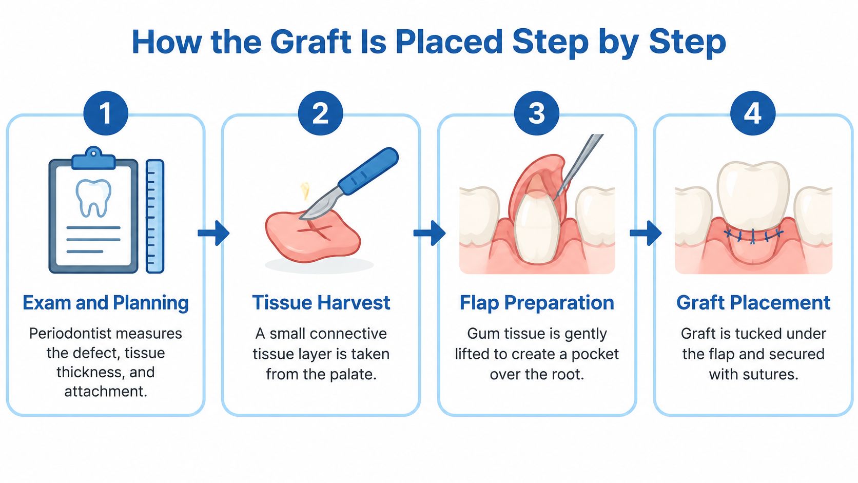
The Procedure in Seven Steps
- Exam and planning. The periodontist measures the defect, checks tissue thickness and attachment, and reviews contributing factors such as brushing pressure or inflammation. When broader anatomy needs review, i-CAT 3D cone beam imaging may support planning.
- Numbing. Local anesthetic is given at both the palate and the gum site. Pressure may still be felt, but sharp pain shouldn't be.
- Donor harvest. A small incision on the palate allows the team to lift connective tissue from beneath the surface lining. The surface layer is preserved where the technique allows, which helps the palate close more cleanly.
- Recipient site preparation. The gum over the exposed root is gently lifted, either as a partial thickness flap or through a tunnel. This creates a pocket where the graft will rest against the root and surrounding tissue.
- Graft placement. The connective tissue is trimmed to size and positioned over the exposed root, beneath the lifted gum. Keeping it flat and still matters, because a graft that shifts doesn't revascularize as reliably.
- Securing the graft. Fine sutures hold the graft in place. In the coronally advanced flap version, the gum is then moved toward the crown and sutured over the graft, covering as much of it as the case allows.
- Protection and instructions. Sutures or a dressing protect the sites, and written aftercare guidance covers eating, brushing, and follow up timing. Patients leave knowing which sensations are expected and which should prompt a call.
Flap or Tunnel
Both designs place the same kind of tissue. They differ in how the gum is opened to receive it. A randomized clinical trial reported in Clinical Oral Investigations found average root coverage of 87% for one approach and 85% for the other, with no statistically significant difference, and complete coverage in 60% and 52% of patients respectively.
That closeness carries a practical lesson. The least invasive design isn't automatically best for every recession pattern. A tunnel may suit several neighboring teeth with intact tissue between them, while a flap may give better access where roots are prominent or more graft volume is needed.
Tissue thickness, interdental support, access, and the size of graft required guide the recommendation. Patients can ask which design is planned and why it fits their defect, because that's a more useful question than asking which technique is newer.
Soreness, swelling, altered sensation, or bleeding can occur during healing. Severity differs from patient to patient, and the recovery section below addresses that trade off directly.
Recovery at the Palate and Gum Site
There's an honest point that belongs at the start of any graft conversation. SCTG creates two healing sites, the gum where the graft sits and the palate where tissue was taken. For many patients, the palate is the site that draws the most attention during the first days.
A 2025 review summarized in PMC identifies the second surgical site, limited graft quantity, lower patient tolerance, and donor site complications as major disadvantages of palatal harvesting. It specifically associated palatal SCTG with more postoperative pain, discomfort, and ulceration because of the extent of the procedure. That's not a reason to avoid the graft outright. It's a reason to weigh the palate as part of the decision, not as a footnote.
Typical Recovery Phases at the Palate and Gum
| Phase | What Patients Often Notice | Practical Focus |
|---|---|---|
| First few days | Soreness, swelling, some bleeding at the palate | Cold compresses, soft foods, and medication as directed |
| First one to two weeks | Tenderness at both sites and healing tissue that may feel different | Gentle care near the surgery and scheduled checks |
| Several weeks | Palate and gum keep healing, and tissue often firms over time | Maintenance cleanings and follow up exams |
| Any time | Slow or uneven palatal healing in some patients | Report unusual pain, bleeding, or ulcers to the office |
Practical care is straightforward but specific. Soft foods in the early days, as directed, keep pressure off both wounds. Brushing stays gentle near the surgery, and other areas are cleaned normally so plaque doesn't build up elsewhere.
Tobacco deserves direct mention because it works against healing and long term stability. Patients who smoke should expect a candid conversation about pausing or quitting around treatment. Follow up visits aren't optional extras either, since sutures, tissue response, and plaque control all need checking.
Avoiding the palate by choosing a biomaterial may reduce recovery burden, but alternatives can yield less thickness or less keratinized tissue, a comparison covered in the graft options section. Comfort, eating limits, and healing time vary, and no responsible plan promises a pain free experience. The periodontist sets the specific instructions for each case.
Realistic Results From Published Studies
Published numbers help, provided they're read correctly. Two terms appear in nearly every study, and they don't mean the same thing. Average root coverage describes how much of the exposed root is covered on average. Complete root coverage means the entire exposed root at a site is fully covered, a stricter bar. A graft can score well on the first measure and still miss the second, the way covering most of a scratch differs from erasing it entirely.
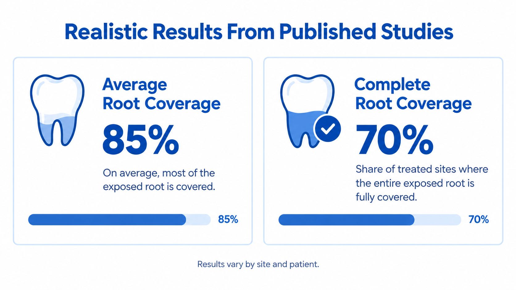
Published SCTG Root Coverage Benchmarks
| Study or Benchmark | Defect Type | Average Root Coverage | Complete Root Coverage |
|---|---|---|---|
| 30-month randomized study, SCTG vs. semilunar coronally positioned flap | Recession defects in the study population | 96.83% (SCTG) vs. 89.25% (comparison) | 88.24% (SCTG) vs. 58.82% (comparison) |
| One-year randomized trial, SCTG alone | Miller Class I and II | Most of each exposed root covered on average | Complete coverage in a majority of patients |
| 2023 Quintessence International study, SCTG control group | Recession defects in the study population | 84.31% ± 17.89% | Not reported as a single rate |
| Systematic review of 23 randomized trials | Varied | Ranged widely across trials, from partial to near-total mean coverage | Ranged from a small minority of sites to nearly all sites |
The spread inside that 23-trial review is the honest part of the picture. Defect classification, tissue thickness, oral hygiene, tooth position, root prominence, and individual healing all move the result. A shallow defect with intact tissue between the teeth carries a different outlook than a deep defect where that support is gone. This is also why the palate donor site stays in the conversation. The coverage numbers are strong enough that many patients accept a second wound, but that choice only makes sense when weighed against the recovery it creates.
Extra steps don't always shift the outcome either. A 12 month randomized trial in Clinical Oral Investigations found average root coverage of 86.0% with 24% EDTA root conditioning and 84.6% without it across 142 recession defects, so that step is individualized rather than assumed necessary.
Recession can also return. Traumatic brushing, ongoing inflammation, tobacco use, and unfavorable tooth position can pull the margin back again. Patients concerned about unsuccessful healing can review information on gum graft failure rates for additional context. Published results describe study groups, and they don't guarantee an individual outcome.
How SCTG Compares With Other Gum Graft Options
Every graft option answers the same question differently: where does the new tissue come from? SCTG borrows it from beneath the surface of the palate, which adds a second wound to recover from. Processed materials skip the palate entirely but generally build less new tissue. Choosing between them means weighing how much coverage and thickness a defect needs against how much healing a patient is willing to take on.
SCTG vs. Other Soft-Tissue Graft Options
| Option | Tissue Source | Root Coverage Evidence | Keratinized Tissue Gain | Best Fit |
|---|---|---|---|---|
| SCTG with coronally advanced flap | Palate, beneath the surface lining | Reference standard; reduced recession more than membranes or processed matrices in pooled trials | Greater gains than enamel matrix or guided regeneration with a flap | Maximum tissue augmentation and predictability |
| Free gingival graft | Palate, surface tissue | Used mainly to build tissue rather than for esthetic root coverage | Widens the band of attached tissue | Adding attached gum where esthetic coverage is not the goal |
| Acellular dermal matrix | Processed donor tissue, no palatal harvest | Comparable for selected Class I and II defects, with SCTG slightly ahead in recession reduction | Gains reported, generally not exceeding SCTG | Avoiding a second surgical site in selected cases |
| Collagen matrix | Processed collagen, no palatal harvest | Percentage root coverage similar to SCTG reported for selected cases | Less keratinized tissue width than SCTG | Selected cases where the palate should be avoided |
| Resorbable guided tissue regeneration | Membrane placed over the defect | SCTG reduced recession more in pooled comparisons | Lower gains than SCTG in reviewed comparisons | Selected defects suited to a membrane approach |
A Cochrane review summarized by the U.S. National Library of Medicine found that SCTG plus a coronally advanced flap reduced recession by an additional 0.37 mm compared with guided tissue regeneration plus a flap, based on three studies involving 98 participants. The same body of evidence reported SCTG advantages of 0.63 mm over acellular dermal matrix and 0.41 mm over resorbable guided tissue regeneration, along with greater keratinized tissue gains than enamel matrix protein plus a flap.
Those advantages are fractions of a millimeter. They carry the most weight when a defect is deep or the existing tissue is thin, and less weight when a small, shallow defect could heal well with either material.
The Verdict by Situation
- Maximum gain matters most. SCTG is often favored when thickness, keratinized tissue, and predictability are the priorities, and the patient accepts a healing period at the palate.
- Avoiding the palate matters most. A collagen matrix or acellular dermal matrix may suit a selected case, accepting that tissue gain may be smaller and recession reduction a fraction of a millimeter less.
- Widening attached tissue is the main goal. A free gingival graft addresses that specific need, usually away from the esthetic zone.
These are evidence trends, not rules for a single mouth. Readers weighing these paths can review gum graft alternatives for a broader overview of materials and what each involves.
One distinction prevents a common mix-up. LANAP laser gum surgery and non surgical therapy treat gum disease and inflammation. They remove infection rather than adding tissue to a receded area, so they answer a different clinical question than grafting does.
Confirm Whether a Graft Fits Your Gums
A consultation gets easier when you arrive with three questions already thought through. Which outcome matters most to you: root coverage, added tissue thickness, comfort, or appearance? Is healing at the palate acceptable, or is avoiding a second surgical site the priority? And are your gum health and plaque control stable enough to support graft healing?
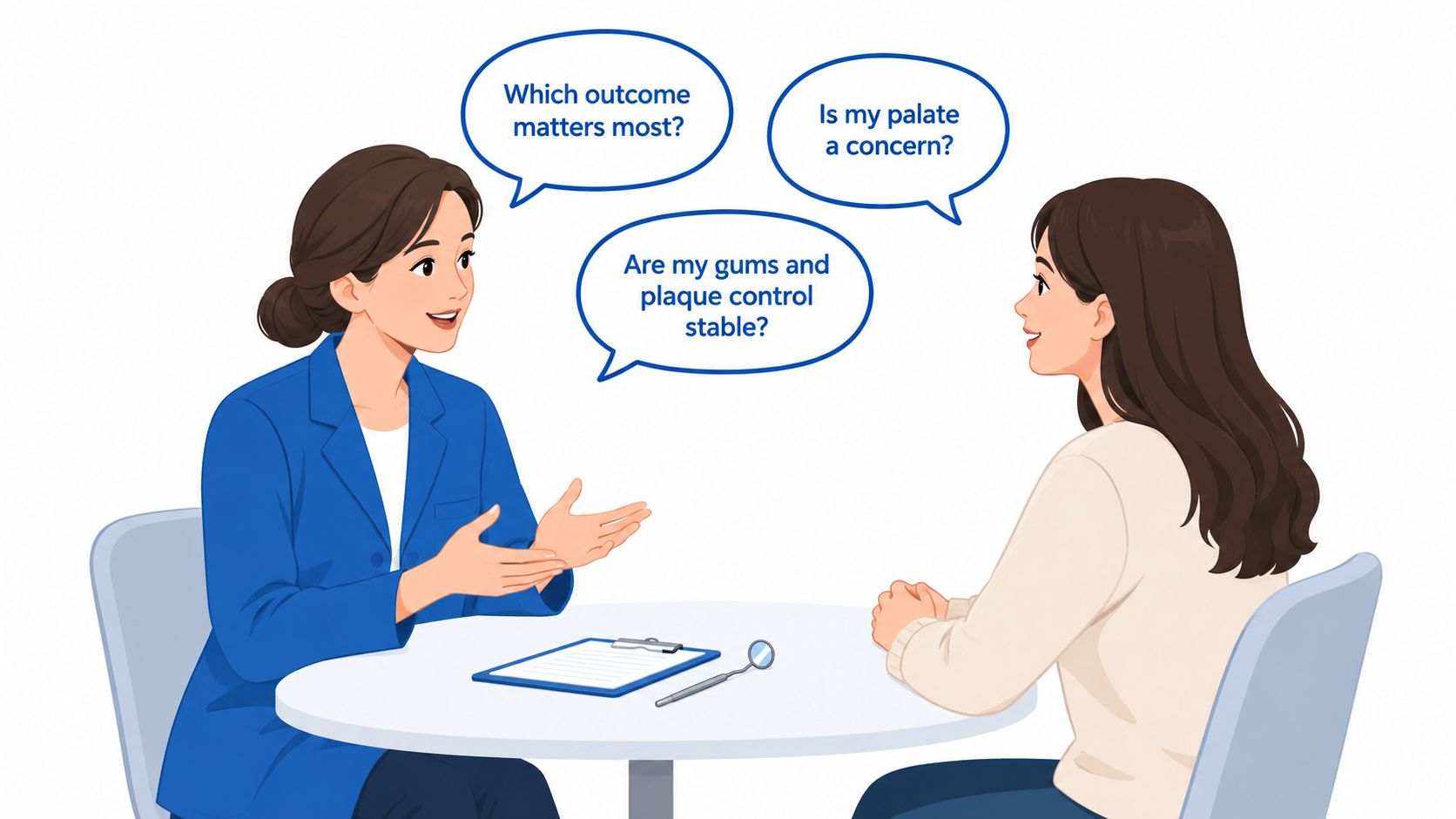
The visit itself usually includes a periodontal exam, measurements of recession depth and gum thickness, and imaging when needed. If active gum disease is present, it gets treated first, since grafting works on stable tissue rather than inflamed gums.
The American Academy of Periodontology explains that gum graft surgery covers exposed roots and may help prevent additional recession and bone loss. Complete root coverage cannot be promised in every case, especially where the supporting tissue between the teeth has already been lost. Bring a written list of questions about palate recovery, expected tissue gain, and other graft options to the exam, so the trade-offs get discussed before anyone schedules surgery.
Periodontal Associates of Delaware provides periodontal evaluation and soft tissue grafting for adults with gum recession. Adults ready for an exam can request an appointment.
Periodontal Associates of Delaware provides board certified periodontal evaluation and soft tissue grafting for adults with gum recession, root sensitivity, and thin gum tissue, with imaging and laser options available when they're clinically appropriate. Patients in Wilmington, Newark, and Dover can visit Periodontal Associates of Delaware to request an appointment at the nearest office and discuss whether a connective tissue graft fits their gums.