Complete root coverage commonly falls below 100%, while average coverage can reach approximately 96%. That means a graft may heal successfully and still leave part of the tooth root exposed.
This distinction matters because many patients search for a gum graft failure rate expecting one clear percentage. Periodontal research doesn't support a single number that applies to every person, tooth, or surgical method. A result may involve excellent healing with partial coverage, complete coverage, early graft loss, later recurrence, or a complication at the gum or donor site.
For adults in Wilmington, Newark, Dover, Pike Creek, Hockessin, Bear, Middletown, and surrounding communities, the useful question isn't just “What is the failure rate?” The more helpful question is, “What outcome is realistic for this specific recession defect, and what factors could affect healing?”
Table of Contents
- Why There Is No Single Gum Graft Failure Rate
- How Recession Type and Tissue Thickness Influence Coverage
- What Normal Healing Looks Like After Gum Graft Surgery
- Beyond Coverage Failure - Other Gum Graft Complications
- Why Recession Can Return After Successful Graft Healing
- Getting an Individualized Assessment from a Delaware Periodontist
Why There Is No Single Gum Graft Failure Rate
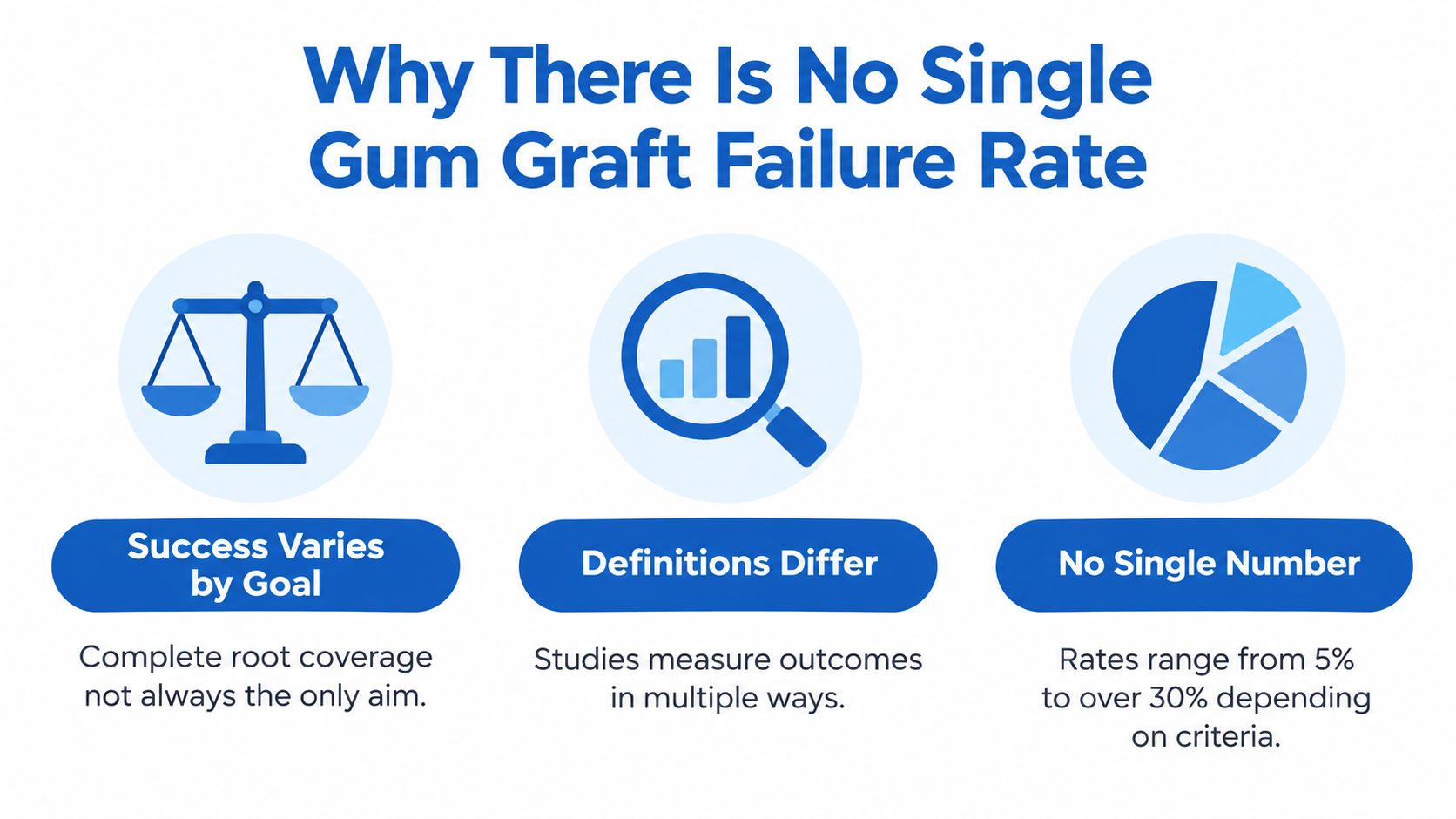
Mean root coverage reached approximately 94% to 98% across four studies, while complete root coverage occurred in 68% to 90% of patients in the three trials that measured it (systematic review of periodontal plastic-surgery procedures). Those figures may seem like a single answer to the question, “What is the gum graft failure rate?” They are not.
A graft outcome can be measured in several ways. The graft may survive and attach, but cover only part of the exposed root. It may provide complete coverage at the final examination. It may heal well and later develop recurrent recession. A patient may also have sensitivity or a donor-site problem even when the graft itself remains attached.
Coverage is not the same as survival
Mean root coverage describes the average portion of exposed root covered across a group of treated teeth. Complete root coverage asks a stricter question: was the entire previously exposed area covered? These measurements are like judging a repaired roof in two ways. One measure asks how much of the roof is protected overall. The other asks whether every opening was sealed.
The same review reported overall mean root coverage of 96% at final examination, but results varied with the procedure, the recession defect, and the study design. A high average can therefore exist alongside some teeth with incomplete coverage. The average describes a group, not a guarantee for one site.
A separate analysis of patients with multiple gingival recessions also found complete coverage in many treated sites, while some sites received partial coverage. Findings from studies of different defects and techniques cannot be placed into one universal percentage. This is why a reported success rate needs its definition, treatment method, and follow-up period.
The goal behind the question
Patients usually want to know whether surgery will protect a root, reduce sensitivity, improve appearance, or help prevent further recession. These goals overlap, but they are not identical. A graft can thicken the gum and provide protection even when a small area of root remains visible. Calling that result total graft loss confuses graft survival with complete coverage.
Patients researching gum graft alternatives may also find different procedures and materials. Their results cannot be compared fairly unless the studies use similar definitions, recession types, follow-up periods, and treatment goals.
At Periodontal Associates, a board-certified periodontist can assess the individual tooth, surrounding tissue, recession pattern, and treatment goal before discussing likely outcomes. The useful question is not only whether a graft “fails.” It is which result is realistic for that site and how success will be measured.
Practical rule: Ask whether a quoted result refers to graft attachment, average root coverage, complete root coverage, appearance, sensitivity, or long-term stability.
No single universal gum graft failure rate applies to every patient, tooth, or technique. A site-specific assessment is more useful than applying a research average to every recession defect.
How Recession Type and Tissue Thickness Influence Coverage
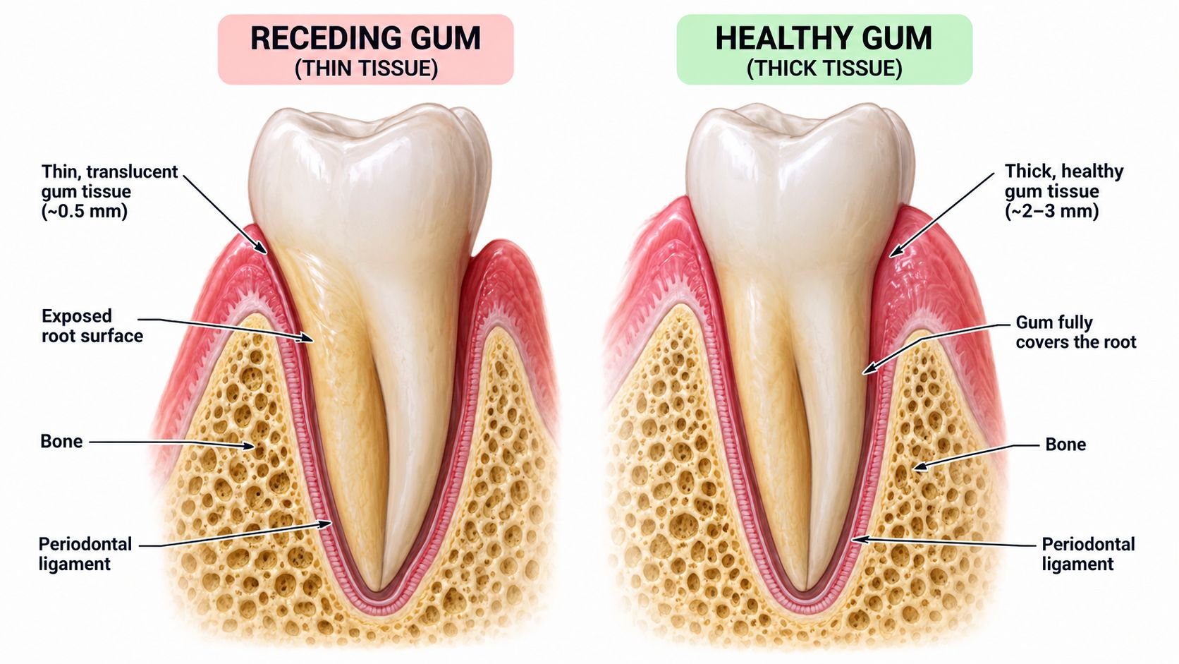
A graft does not heal in isolation. The gum around the tooth must provide blood supply, stable support, and enough tissue to cover the exposed root without excess tension. If those conditions are limited, the graft may survive and strengthen the area while still leaving part of the root visible. That outcome is partial coverage, not the same as complete graft loss.
Periodontists classify recession by examining the defect and the tissue between neighboring teeth. Miller Class I and II defects may achieve full coverage because the supporting tissue between the teeth remains largely intact. Class III defects often allow partial coverage because loss of interdental bone or attachment limits how far the gum can be moved. Class IV defects usually have little potential for surgical root coverage, as described in this British Dental Journal review of recession treatment.
Why the tissue phenotype matters
Gum tissue may be relatively thick or thin, a difference often called the gingival phenotype. Thick tissue provides more substance for healing and for moving the gum toward the crown of the tooth. Thin tissue offers less protection from tension, movement, brushing trauma, and future recession.
A systematic review found mean root coverage of about 55% to 85% for Miller Class III and Cairo RT2 defects, while complete root coverage occurred in about 38% of cases. The same review found that a thick gingival phenotype was associated with a higher likelihood of complete coverage, with an adjusted relative risk of 1.52 compared with a thin phenotype. It also reported complete root coverage of 80.0% versus 48.8% when comparing connective-tissue grafts with acellular dermal matrix in selected cases. These findings come from a systematic review of recession predictors and graft materials, and they describe study groups, not a guaranteed result for every tooth.
Several local and patient factors can change the prognosis:
- Interdental bone and attachment: Reduced support between teeth limits how much tissue can move over the root.
- Keratinized-tissue width: A narrow band of firm gum may provide less stability during healing.
- Root prominence: A prominent root can make close adaptation and immobilization more difficult.
- Recession height and width: Larger or deeper defects present a greater technical challenge.
- Plaque control and inflammation: Inflamed tissue creates a poorer healing environment than controlled periodontal tissue.
- Smoking and nicotine exposure: These exposures can interfere with periodontal surgical healing, so nicotine use should be included in the risk assessment.
Why the examination must come first
Before surgery, a periodontist should record the recession classification, interproximal attachment, tissue thickness, keratinized-tissue width, root shape, smoking or nicotine status, plaque control, and brushing habits. Those findings help determine whether a connective-tissue graft fits the site or whether the surgical design needs modification.
At Periodontal Associates, a board-certified periodontist can assess these features tooth by tooth. That site-specific review is more useful than applying one generic gum graft failure rate to every recession. The expected result may involve graft survival, partial coverage, complete coverage, or protection with some root exposure remaining.
What Normal Healing Looks Like After Gum Graft Surgery
The first postoperative week can create considerable anxiety because the graft may not look like ordinary pink gum tissue. Early whitening or a pale appearance can be part of normal healing. Appearance alone can't confirm success or failure, especially before the tissue has matured.
A progressively dark or dusky graft deserves prompt attention. The same is true for visible separation, increasing pain, pus, foul taste, or persistent bleeding. Those signs may indicate compromised integration, infection, or another healing problem and should be reported to the treating periodontist rather than watched at home.
A simple way to judge changes
Patients can focus on the direction of symptoms instead of expecting the site to look normal immediately.
- Color: Early whitening or paleness may occur, but progressive darkening or a dusky appearance requires professional review.
- Pain: Some soreness can occur after surgery. Increasing pain, particularly when it worsens instead of settling, should be reported.
- Stability: The graft should remain protected and undisturbed. Visible separation or movement isn't a normal reason to wait.
- Drainage or taste: Pus, a foul taste, or a bad odor may signal infection.
- Bleeding: Persistent bleeding needs assessment, especially when pressure and the prescribed instructions don't control it.
A patient shouldn't pull at the graft, repeatedly inspect it with a finger, brush directly over it, or test whether it has attached. Mechanical disturbance can disrupt the delicate blood supply. Tobacco and nicotine can also interfere with healing, while plaque, pressure, movement, and local inflammation may compromise integration.
Healing depends on blood supply and stability. Protecting the graft matters more than searching for a supplement or rinse that promises to change the outcome.
Postoperative instructions vary with the procedure and the location treated. The periodontist may adjust oral hygiene, diet, activity, and rinsing instructions to protect the surgical site. A patient who has also received treatment for a periodontal defect may need coordinated care, including evaluation of the supporting bone. Information about periodontal bone grafts can help explain why hard-tissue and soft-tissue problems sometimes require separate assessments.
A graft that looks unusual doesn't automatically mean it has failed. Early assessment is still the safest choice when symptoms are worsening. The periodontist may determine that the tissue is healing normally, that only part of the graft integrated, or that infection or another complication needs treatment. A second procedure, if appropriate, is considered only after the area has matured and the original cause has been evaluated.
Beyond Coverage Failure - Other Gum Graft Complications
A result can be unfavorable without fitting the simple image of a graft that “didn't take.” Incomplete root coverage is one possibility, but healing problems can also involve the recipient site, the donor site, or the surrounding tooth and bone.
Reported unusual recipient-site complications have included bone exostosis, epithelial inclusion, root resorption, abscess, tissue overgrowth, liver-clot formation, and oroantral communication. A systematic review examined 1,434 articles and included 28 studies, consisting of one randomized trial, two cohort studies, and 25 case reports or case series (systematic review of unusual recipient-site complications).
Why these reports need careful interpretation
Bone exostosis and epithelial inclusions were among the most frequently reported unusual complications after subepithelial connective-tissue grafts, free gingival grafts, and acellular dermal-matrix allografts. However, much of the evidence came from case reports and case series. Those reports can identify problems that clinicians need to recognize, but they don't establish a routine percentage risk for every patient.
That distinction prevents two common errors. A patient shouldn't assume that every listed complication is likely, and a practice shouldn't describe grafting as a risk-free procedure. The appropriate risk discussion depends on the procedure, the number of sites, the patient's health, the tissue anatomy, and the findings during follow-up.
What follow-up helps identify
A follow-up visit can distinguish among several different situations:
- Incomplete coverage: The graft healed but didn't cover the entire exposed root.
- Wound-healing problem: The tissue is not progressing as expected or has become unstable.
- Infection or abscess: Drainage, worsening pain, swelling, or an unpleasant taste may require prompt treatment.
- Donor-site symptoms: The palate may remain uncomfortable or heal more slowly than expected.
- Tissue change: Overgrowth, an epithelial inclusion, or another localized change may need evaluation.
- Tooth or bone involvement: Root resorption, bone changes, or an oroantral communication require a different clinical response.
Multiple recession sites can make recovery harder to assess because each location may heal differently. Individual postoperative instructions and scheduled review allow the periodontist to respond to the actual pattern rather than labeling every difference as failure.
Patients with swelling, pus, or an unpleasant taste should seek prompt periodontal advice, including evaluation for a periodontal gum abscess. Early contact doesn't mean the graft has been lost. It gives the clinician an opportunity to identify the cause and protect the healing tissues.
Why Recession Can Return After Successful Graft Healing
Early graft failure and later recurrence describe different timelines. Early failure involves a problem with survival, attachment, or healing soon after surgery. Recurrence develops later, after the area initially appeared stable, when recession begins to return.
A graft adds a layer of protective tissue, but it may not remove every cause that stressed the gum in the first place. Traumatic toothbrushing, plaque buildup, periodontal inflammation, tooth position, and unusual biting forces can continue affecting the site. For example, repeatedly scrubbing a previously vulnerable area can gradually injure the new tissue, much like rubbing a healing patch before it has fully strengthened.
Long-term protection requires behavior and maintenance
Gentle oral hygiene still requires consistent cleaning. The goal is to follow the periodontist's instructions without pressing, scrubbing, or repeatedly traumatizing the graft. Inflammation control matters because active periodontal disease can weaken the supporting tissues around the tooth.
A long-term plan may include:
- Technique review: A dental professional can demonstrate how to clean the area with enough care to remove plaque without excessive force.
- Periodontal maintenance: Regular monitoring can identify inflammation, tissue thinning, or new recession while the change is still limited.
- Occlusal evaluation: Unusual biting forces or tooth contacts may need attention if they place repeated stress on the tissue.
- Smoking cessation: Avoiding tobacco supports the blood supply and the environment needed for healing and stability.
- Cause control: Tooth position, a strong frenum pull, plaque retention, or another local factor may require treatment.
The palate is part of the outcome
Connective tissue taken from the palate creates a second healing site. A 2024 narrative review linked palatal donor-site complications with harvesting technique and reported bleeding rates ranging from 1.2% to 33% (narrative review of connective-tissue graft complications). That range describes results across different procedures, studies, and patient groups, not a personal prediction.
Excessive undermining of the palatal flap can reduce its blood supply, delay healing, or contribute to tissue necrosis. Donor-site protection and clear postoperative instructions therefore matter alongside care of the grafted area.
A recurrent site needs a site-specific examination rather than a generic failure percentage. At Periodontal Associates of Delaware, the periodontist may compare earlier and current photographs, measure recession and tissue thickness, check for bleeding or inflammation, review brushing technique and nicotine exposure, and assess tooth position and biting forces. The examination helps identify which factor is active now and whether treatment should focus on behavior, inflammation, bite forces, the local anatomy, or another cause.
Getting an Individualized Assessment from a Delaware Periodontist
A generic gum graft failure rate can't show how a particular tooth will respond. A useful evaluation measures the factors that shape coverage, including tissue thickness, keratinized-tissue width, clinical attachment level, recession height and width, tooth type, root prominence, interproximal support, plaque control, and smoking or nicotine exposure.
A randomized clinical-trial analysis of tunnel surgery with subepithelial connective-tissue grafts for multiple recessions found that baseline measurements predicted later results. Each 1-millimeter increase in baseline clinical attachment level was associated with higher odds of failing to reach more than 85% mean root coverage or complete root coverage. Each 1-millimeter increase in baseline keratinized-tissue width improved the likelihood of achieving more than 85% mean root coverage, complete root coverage, and the highest reported esthetic score (clinical-trial analysis of baseline predictors).
What the consultation should clarify
A board-certified periodontist can help a patient understand the difference between a realistic goal and an ideal result. The discussion may include:
- The reason for treatment: Root sensitivity, protection of an exposed root, progressive recession, tissue thickening, or esthetic concerns.
- The defect classification: The amount of supporting tissue between teeth and the severity of recession affect predictability.
- The tissue phenotype: Thin tissue may need a different approach from thick tissue.
- The surgical option: A connective-tissue graft, another soft-tissue approach, or observation may be considered based on the anatomy.
- The endpoint: The patient should know whether the goal is complete coverage, meaningful partial coverage, increased tissue thickness, or improved comfort.
- The maintenance plan: The cause of recession must be managed so that the result has a better chance of remaining stable.
The evaluation may also identify periodontal disease or bone loss that needs attention before soft-tissue surgery. LANAP laser gum surgery using the FDA-cleared PerioLase MVP-7 may be considered for appropriate periodontal disease treatment, but it isn't a substitute for a recession evaluation or a guarantee of root coverage. Soft-tissue grafting and periodontal disease control address different clinical problems, although both may matter in the same patient.
How imaging supports broader planning
Soft-tissue recession is assessed directly through a periodontal examination. When bone volume, tooth position, or implant planning is also involved, i-CAT 3D cone beam imaging can provide three-dimensional information about the supporting structures. X-Nav dynamic surgical navigation may support implant planning and placement when a missing tooth and inadequate bone are part of the treatment picture.
Periodontal Associates of Delaware provides periodontal and implant care through board-certified periodontists at offices in Wilmington, Newark, and Dover, serving adults across New Castle and Kent Counties. The practice offers soft-tissue grafting, periodontal treatment, LANAP with the PerioLase MVP-7, i-CAT imaging, and X-Nav navigation when clinically appropriate.
A consultation shouldn't be used to promise a particular percentage. It should help the patient understand the condition of the tissue, the limits of coverage, the likely risks, and the steps that may improve healing. Individual results vary, and readers should consult a periodontist for site-specific guidance rather than relying on an internet-wide gum graft failure rate.
If gum recession, exposed roots, sensitivity, bone loss, or missing teeth are causing concern, patients can request an evaluation at the nearest Wilmington, Newark, or Dover office. Periodontal Associates of Delaware offers board-certified periodontal assessment, soft-tissue grafting, periodontal disease treatment, and implant planning matched to each patient's anatomy and goals. Request an appointment at the most convenient location through the appointment request page.