The extraction appointment is approaching, and the treatment plan includes an unfamiliar term: tooth extraction membrane. For someone already concerned about losing a tooth, another material or procedure can raise understandable questions about healing, necessity, and what happens next.
A membrane isn't automatically needed after every extraction. Its value depends on the remaining bone, the condition of the socket, and the plan for replacing the tooth. Understanding those differences can make the treatment discussion feel more manageable.
Table of Contents
- What Is a Tooth Extraction Membrane
- Why the Jawbone Shrinks After Extraction
- Resorbable vs Non-Resorbable Membranes
- How a Membrane Is Used in Socket Preservation
- What the Research Shows About Membrane Benefits
- Does Every Extraction Socket Need a Membrane
- What to Expect During Healing and Recovery
- How Membrane Choice Affects Future Implants
- Questions to Ask Your Periodontist
What Is a Tooth Extraction Membrane
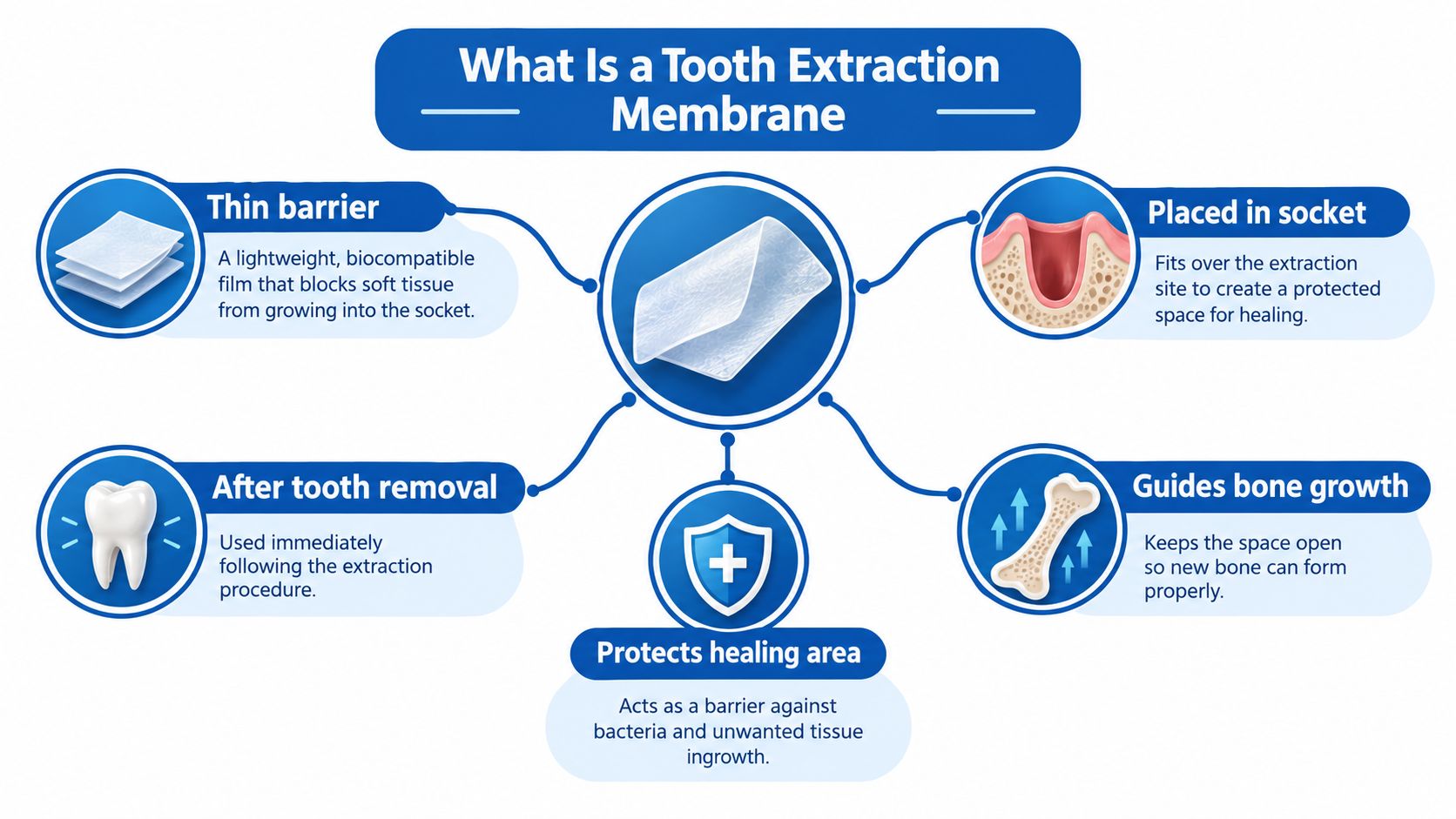
A patient may expect an extraction to involve removing the tooth and letting the opening heal. Hearing that a thin barrier may also be placed can sound more complicated than it is. A tooth extraction membrane is a protective layer used over or around the healing socket, the space left behind after removal.
Its purpose is to help organize healing rather than replace the missing tooth. Membranes are used in socket preservation, which aims to maintain the shape of the tooth-supporting ridge, and in guided bone regeneration, which protects an area where bone formation is needed. They help separate the healing bone space from surrounding soft tissue, as described in a systematic review of membrane use after extraction.
A membrane and a bone graft have different jobs
Patients often hear these terms together, but they don't describe the same material.
- Bone graft material occupies space within the socket and supports the planned reconstruction.
- A membrane forms a barrier around the area being protected.
- Socket preservation describes the overall treatment approach, which may involve a graft, a membrane, or a selected combination.
A useful comparison is a protected garden bed: the material inside the bed and the covering over it serve different purposes. Neither should be selected because the other is being used.
Practical distinction: Agreeing to bone grafting doesn't automatically mean that every socket needs a membrane.
The important question is what the barrier would contribute in that particular site. A treatment explanation should identify the problem being addressed, not just name the material being placed.
Why the Jawbone Shrinks After Extraction
A patient can leave an extraction appointment with a neat stitch, an easy recovery, and a gumline that looks healed within a month, then hear at an implant consult that the ridge has “collapsed.” The surprise is understandable. The opening can close while the bony wall that used to hold the tooth becomes thinner.
The reason is mechanical as much as biological. A tooth is not sitting in inert concrete; the socket wall, especially the cheek-side plate, is maintained by the tooth and its ligament. Remove the tooth and the body recycles part of that thin support, similar to a retaining wall losing the post that braced it. Human studies of untreated sites report about 2.6–4.6 mm of ridge-width loss and 0.4–3.9 mm of height loss, with the facial side often affected most, in research on post-extraction ridge changes.
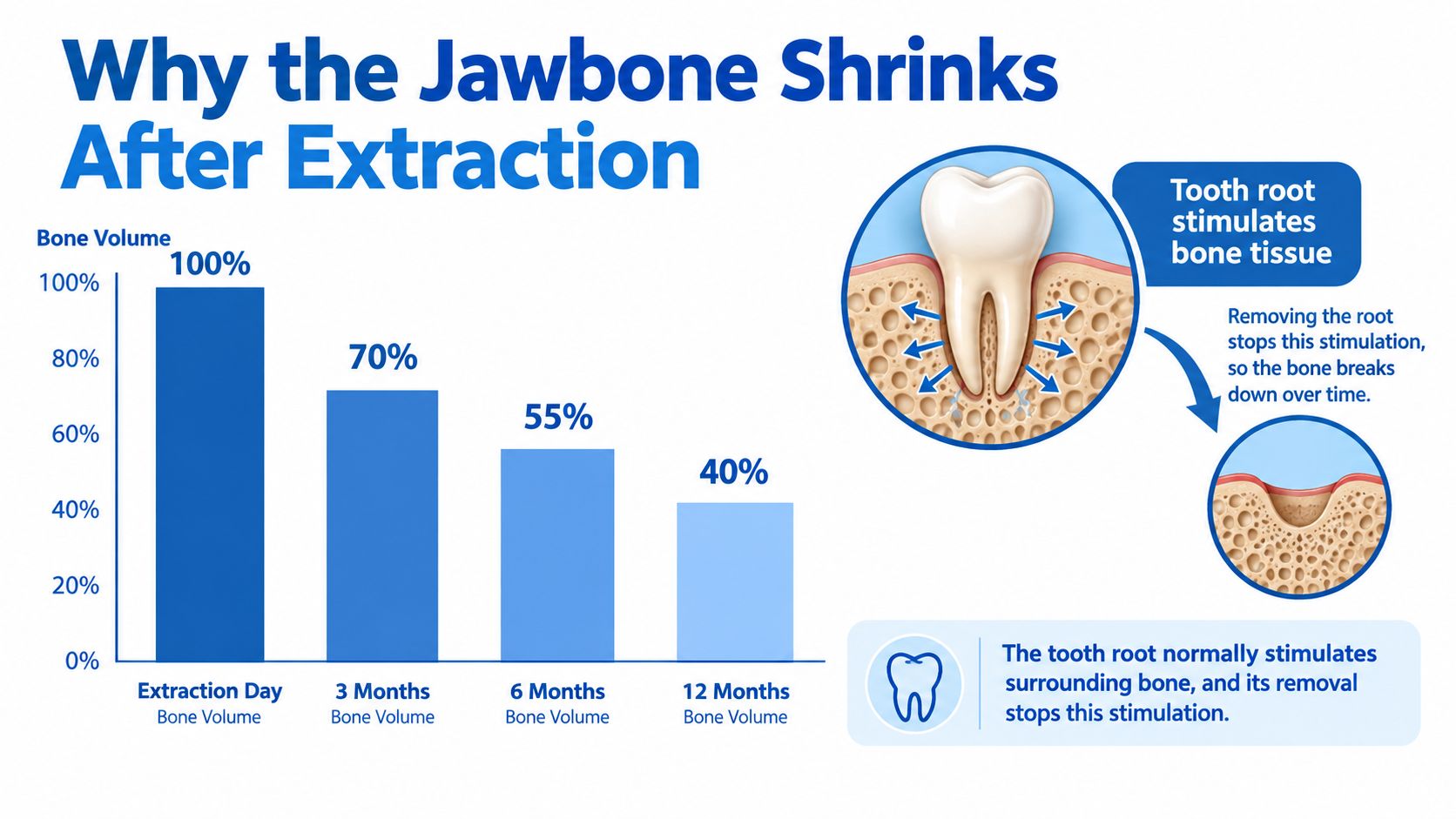
The illustration is a conceptual aid rather than a patient-specific forecast. It should not be read as a fixed percentage timetable for your jaw.
Confusion usually comes from mixing two healing events. The socket can fill with new bone in the middle while the outer outline still narrows, the way a room can be refurnished after one wall is moved inward. That is why a site may look tidy at the gum surface yet measure differently underneath.
A membrane enters the discussion only if contour has a job to do. Width influences implant position, height affects available support, and cheek-side shape influences how the gum drapes around a future tooth. If none of those goals are threatened, remodeled bone may still be acceptable. If they are, preserving space early can be more predictable than rebuilding it later.
Resorbable vs Non-Resorbable Membranes
Resorbable membranes break down in the body. Non-resorbable membranes don't dissolve and require a plan for later removal. These categories describe how the barrier behaves, not a ranking from better to worse.
The choice should match the defect, the needed barrier function, and the way the gum tissue will be managed. A material that suits an enclosed socket may not be the preferred option for an area with missing bone walls.
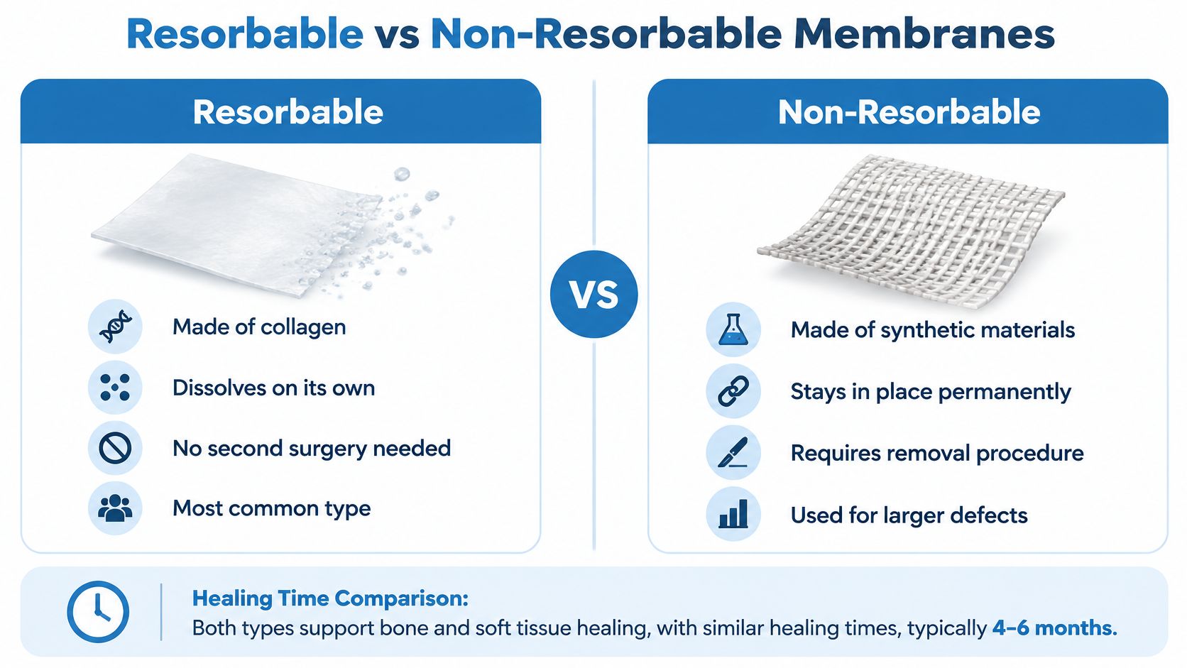
The graphic's reference to a non-resorbable membrane remaining permanently means that it doesn't dissolve on its own, not that it should routinely remain indefinitely. The treating periodontist determines its removal plan.
| Patient concern | Resorbable membrane | Non-resorbable membrane |
|---|---|---|
| Typical material | Commonly collagen | Materials such as dense or expanded PTFE |
| What happens afterward | Breaks down over time | Remains until removed |
| Removal planning | Routine retrieval generally isn't needed | A removal appointment or procedure is planned |
| Selection considerations | Tissue integration and resorption behavior | Duration of protection and exposure management |
Material details matter
Not all collagen membranes behave alike. Processing and structure affect how they integrate with tissue and maintain their barrier function. A systematic review of collagen membranes found results favoring non-crosslinked over crosslinked membranes in its analysis, but also emphasized variation among clinical situations.
That finding doesn't mean patients should select a product category without an examination. A periodontist must consider whether the membrane can remain stable, whether gum coverage is required, and how the local defect will be protected.
A resorbable membrane also doesn't eliminate follow-up visits. Even when no retrieval procedure is expected, the clinician still needs to check the tissue and confirm that healing is progressing appropriately.
How a Membrane Is Used in Socket Preservation
Socket preservation begins with careful extraction, not with opening a package of graft material. The periodontist aims to protect the remaining socket walls, assess the defect, and choose an approach that fits what is present.
The final plan may change after the tooth is removed because the socket can then be examined directly. The membrane is one part of that plan, not a substitute for proper cleaning or stable wound management.
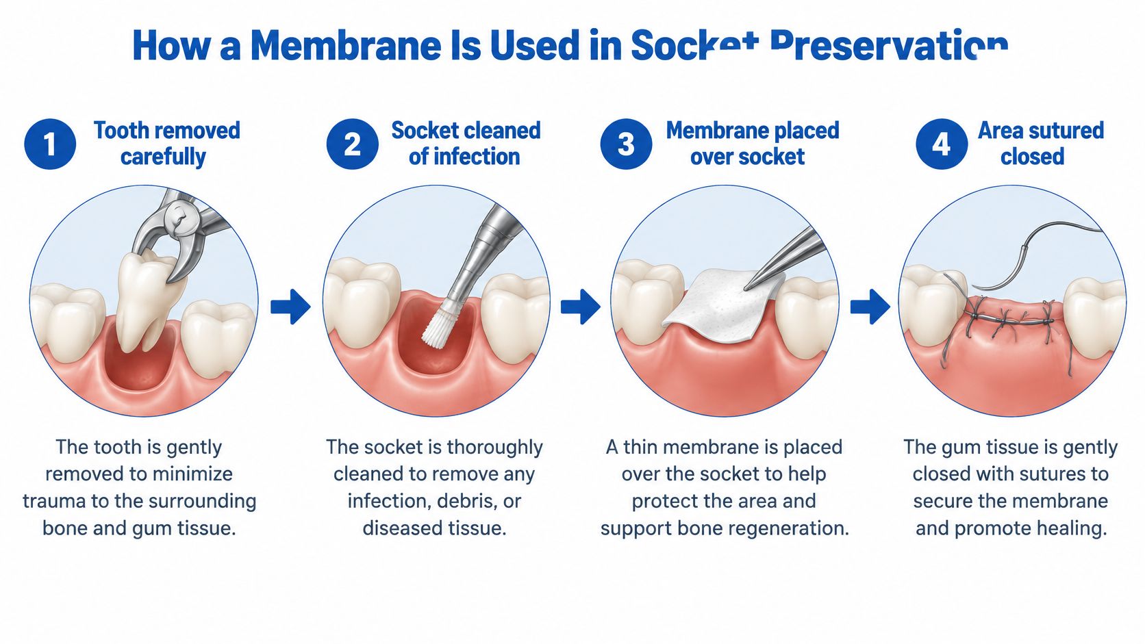
What happens at the treatment site
- Careful removal and assessment. The clinician removes the tooth while trying to preserve the surrounding structures, then examines the remaining walls.
- Socket cleaning. Infected or inflamed granulation tissue is removed so the site can be evaluated and treated.
- Grafting when indicated. The selected material is placed according to the defect and future restoration plan. The practice's explanation of periodontal bone grafts provides related background.
- Barrier placement and stabilization. When indicated, the membrane is positioned to protect the site and help contain graft material. Stitches may stabilize the membrane, the surrounding gum tissue, or both.
The illustration simplifies closure. Not every membrane is completely covered by gum tissue; some selected techniques involve intentional exposure.
A successful protocol depends on how the site is prepared and stabilized, not simply on whether a membrane is present.
Evidence supporting the combined approach includes a randomized study of bovine bone mineral with a collagen membrane. During the first four months, ridge width decreased by 1.04 ± 1.08 mm with preservation versus 4.48 ± 0.65 mm after extraction alone.
Because that study tested a combined treatment, its results shouldn't be attributed to the membrane alone. The graft, barrier, socket condition, and surgical management all belong in the explanation.
What the Research Shows About Membrane Benefits
A patient may hear “we'll place a membrane” and assume the ridge is now protected from all shrinkage. The research is more measured: membranes can improve preservation odds, especially as part of a planned protocol, and the final result still depends on socket walls, blood supply, flap closure, and healing.
A membrane works like temporary fencing around a construction site. It helps keep the right cells in and the fast-growing gum tissue out, yet it cannot rebuild a wall that is already missing.
In a systematic review of barrier membranes used alone, treated sites averaged 0.909 mm more bone height and 2.966 mm more ridge width than extraction alone. Those are pooled averages across different patients and defects, useful for understanding direction of effect rather than predicting one person's result.
Study conclusions vary because studies do not start with identical mouths. A systematic review and meta-analysis of socket-sealing materials included 12 clinical trials, 321 treated sockets, and 312 sites available for analysis; it reported a statistically significant horizontal-preservation advantage for resorbable membranes over no membrane, while substantial variation kept the authors from naming one universally superior method.
Evidence is still developing. A 2024 review of bone-mineral grafts with resorbable collagen barriers found greater ridge preservation overall, said the trend did not reach statistical significance, and called for more clinical studies.
If an edge becomes exposed, call the office instead of trimming or pulling it. Management can range from monitoring to adjustment, depending on the material and tissue response.
The practical reading: membranes are evidence-based tools whose value depends on anatomy, and the research does not support treating every socket identically.
Does Every Extraction Socket Need a Membrane
No. Membrane selection should be anatomy-dependent, not automatic. A recommendation should explain why the barrier adds value in that particular socket.
Consider the difference between an extraction site with thick, intact cheek-side bone and one with a thin or missing outer wall. Those sites present different challenges, even if both patients plan to replace the missing tooth with an implant.
A randomized clinical trial in sites with thick buccal bone followed 20 participants for six months and found comparable measured outcomes with collagenated bovine graft alone and the same graft covered by a non-crosslinked collagen membrane. The authors noted that omission could reduce treatment time, cost, and additional materials in that clinical setting.
What that finding does and doesn't mean
The result supports a selective approach. It doesn't establish that membranes are unnecessary in all extraction sites or that patients should decline a recommended barrier without understanding the defect.
| Clinical feature | Question for treatment planning |
|---|---|
| Thick, intact cheek-side bone | Would a membrane add meaningful protection beyond the selected graft? |
| Thin or damaged socket walls | Is additional containment or protected space needed? |
| Limited soft-tissue coverage | Can the selected barrier be managed predictably? |
| Infection or medical risk factors | Does the site require a different approach or closer reassessment? |
The trial's findings shouldn't be generalized to large defects, thin cheek-side bone, compromised coverage, infected sites, or patients with systemic risk factors.
More biomaterials don't necessarily produce a better result in every socket.
The strongest recommendation is the one that connects the material choice to a clearly identified clinical need. Sometimes that means using a membrane. In favorable anatomy, a simpler approach may be reasonable.
What to Expect During Healing and Recovery
The most useful recovery instructions are specific to the membrane and closure technique used. Patients should leave knowing whether any barrier is expected to remain visible, whether removal is planned, and whom to contact if the site changes.
Tenderness, swelling, and stitches may be part of recovery, but symptoms should be interpreted in context rather than against a rigid online timeline. The practice's guidance on gum surgery stitches can help clarify questions about sutures and follow-up.
When the care team should be contacted
- Increasing pain or swelling. Worsening symptoms, particularly after initial improvement, warrant prompt review.
- Unusual discharge or a persistent unpleasant taste. These changes should be reported rather than managed with leftover medication.
- A visible or moving membrane. The periodontist should determine whether this is expected exposure or an unplanned change.
- Persistent material at the site. Patients shouldn't pull, trim, or attempt to remove suspected membrane remnants.
- Severe symptoms. Uncontrolled bleeding or difficulty breathing or swallowing requires urgent care.
Exposure doesn't automatically mean treatment failure. Some barriers are intentionally managed with exposure, while unexpected exposure requires assessment of stability, tissue condition, and possible infection. Depending on those findings, the clinician may recommend observation, antimicrobial care, removal, or revision.
Health history affects the plan
Smoking, diabetes, and glycemic control belong in the preoperative discussion. Patients should disclose medical conditions and medications so the periodontist can tailor counseling and monitoring.
A clinical study of microscope-assisted ridge preservation illustrates why early appearance alone isn't enough to judge the outcome: improved early visual healing didn't translate into significant differences in crestal bone healing or patient-reported outcomes at 12 weeks.
Evidence for medically complex patients remains less settled. A reassuring-looking surface doesn't replace clinical follow-up, and a concerning appearance doesn't establish failure without an examination.
How Membrane Choice Affects Future Implants
A socket can heal well and still leave bone in the wrong place. One common scenario: the ridge looks full at the surface, but imaging shows it narrows exactly where an implant body would need to sit. Preserved bone only counts if it ends up where the replacement tooth will go.
Implant planning therefore runs backward from the final crown. The periodontist first marks where the new tooth must sit for biting, appearance, and gum support, then maps the implant position beneath it, then checks whether the bone can hold that position. A house works the same way: the floor plan comes first, and the foundation is poured to match it. A membrane selected with that plan in mind protects a defined volume where the implant will need it.
The checkpoints run on a timeline
Before extraction, the clinician weighs whether preservation fits the intended replacement at all, since a socket headed for a bridge or denture may call for a different approach. During healing, follow-up confirms the protected site is maturing as expected. Before implant placement, a fresh evaluation measures what the bone delivered.
Three-dimensional imaging sharpens each of those checks. At the practice's Wilmington, Newark, and Dover offices, implant planning can include i-CAT 3D cone beam imaging and X-Nav dynamic surgical navigation where appropriate. These tools improve assessment and surgical precision; they neither shorten biological healing nor guarantee a result. Patients weighing an implant can review the practice's guide to the space and bone an implant needs to see how crown position, implant placement, and neighboring structures relate.
Even careful preservation can be followed by additional grafting, and available evidence has not established that ridge preservation reliably removes that possibility. A sound plan says so plainly. If healing delivers less bone than expected, the periodontist should already have outlined the fallback, whether that means staged augmentation or adjusted timing.
For adults across New Castle and Kent Counties, including Pike Creek, Hockessin, Bear, Middletown, and surrounding communities, the periodontal team coordinates with the restorative dentist so each step serves the planned tooth.
The membrane is one decision inside a longer sequence.
Questions to Ask Your Periodontist
A consultation should turn unfamiliar terminology into a clear explanation of the site, the options, and the follow-up plan. Patients don't need to choose a membrane themselves. They should understand why the clinician recommends one approach over another.
The most useful questions focus on the reason for treatment rather than the name of a product. A written treatment plan can help patients compare what is essential, what is optional, and what remains uncertain until extraction.
A consultation checklist
- Need for extraction. Can the tooth reasonably be retained, or is removal the recommended course?
- Socket anatomy. Is the cheek-side bone thick, thin, damaged, or missing?
- Purpose of the barrier. What specific benefit is expected from a membrane in this site?
- Alternatives. Could grafting alone or another healing approach be appropriate?
- Material selection. Is the proposed membrane resorbable, and what follow-up or removal will it require?
- Coverage and exposure. Will the barrier be covered, or is some visibility expected?
- Recovery planning. Which changes require a call, and how will concerns be assessed?
- Future treatment. What must be confirmed before implant placement, and could additional grafting still be needed?
The central decision
A tooth extraction membrane is neither an automatic requirement nor an unnecessary extra in every case. Its usefulness depends on the defect, the intended restoration, and the conditions needed to protect healing.
For patients whose main concern is gum disease rather than an unavoidable extraction, the discussion may also include tooth-preserving treatment options. These can include LANAP laser gum surgery using the FDA-cleared PerioLase MVP-7 when clinically appropriate; that is a separate decision from socket preservation.
Individual results vary, and readers should consult a periodontist for advice based on an examination, health history, and appropriate imaging.
The board-certified periodontal team at Periodontal Associates of Delaware offers extraction site preservation, bone grafting, and coordinated implant planning, with membrane selection based on each patient's anatomy and treatment needs. Patients can request an appointment at the nearest Wilmington, Newark, or Dover office to review whether a tooth extraction membrane belongs in their care plan.