A periodontist has just explained that a failing tooth may need removal, followed by a bone graft. The word “graft” can make an ordinary dental problem sound like major reconstructive surgery. Many patients also wonder whether the material comes from another person, whether the procedure will hurt, and whether the graft is truly necessary.
A periodontal bone graft is better understood as foundation repair. Gum disease, tooth loss, infection, or extraction can leave too little bone to support a natural tooth or future implant. The right graft material and technique depend on the defect, the location, the condition of the surrounding tissue, and the long-term restoration plan. Individual results vary, so a periodontist should evaluate each case before treatment is chosen.
Table of Contents
- Why Bone Grafting Is a Common Part of Gum Care
- Understanding the Four Main Types of Bone Grafts
- When You Need Ridge Preservation or Bone Augmentation
- The Healing Process and What to Expect After Grafting
- Addressing Common Concerns About Pain and Risks
- Next Steps for Patients in Wilmington, Newark, and Dover
Why Bone Grafting Is a Common Part of Gum Care
A patient with advanced gum disease may still have a tooth that looks reasonably normal above the gumline. Underneath, however, infection may have created a pocket and removed part of the bone that once held the root firmly. Another patient may have lost a tooth years earlier and now have a narrow ridge that cannot safely support an implant. In both situations, the visible concern is the tooth or missing space, but the treatment challenge is often the underlying bone.
Bone grafting isn't a recent experiment. A review of periodontal therapy traces the use of grafts to rebuild bone defects back to Hegedus in 1923, making the concept roughly a century old. Modern periodontal regenerative and reconstructive procedures have developed far beyond those early operations. A review of bone grafts in periodontics describes better long-term clinical outcomes for regenerative treatment in intrabony defects than for open flap debridement alone in some evidence, including shallower residual probing depths and greater clinical attachment gains.
The foundation matters more than the label
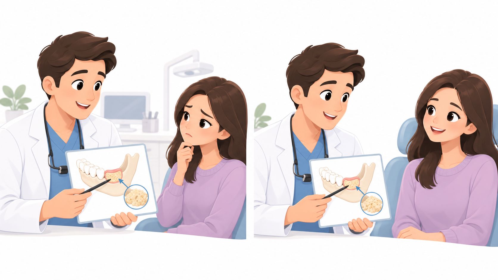
A graft doesn't “fill a hole” and instantly become normal bone. It usually acts as a scaffold or space maintainer, giving the body a stable area where bone-forming cells can work. The periodontist may use it to support remaining teeth, preserve an extraction socket, rebuild a deficient ridge, or prepare for implant placement.
The recommendation can feel surprising when gum recession is the main visible problem. Recession affects the soft tissue covering the root, while bone loss concerns the hard tissue supporting the tooth. These problems can occur together, but they aren't treated in exactly the same way. Patients with recession can review information about bottom gum recession and its treatment options while the periodontist determines whether bone grafting, soft tissue grafting, periodontal therapy, or a combination is appropriate.
Practical rule: The important question isn't simply whether a graft is available. It's whether the defect has the shape, blood supply, tissue support, and treatment goal that make grafting useful.
The evidence also calls for realistic expectations. Regeneration can improve measurable clinical findings, but it doesn't promise a return to the exact bone anatomy present before gum disease or extraction. A careful plan separates disease control from reconstruction and explains what the graft can and cannot accomplish.
Understanding the Four Main Types of Bone Grafts
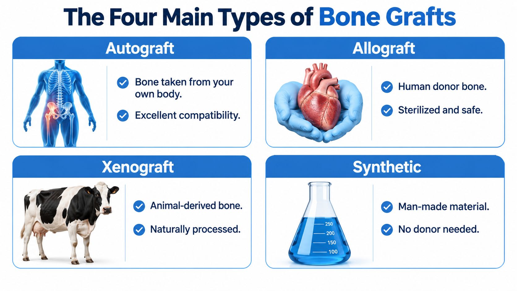
The four main graft categories differ by source and behavior. The material choice isn't a popularity contest. A periodontist selects the option that fits the defect's size and shape, the need for volume stability, the desired healing pattern, and the patient's preferences.
A contemporary review reported that, in the United States, mineralized allografts account for 37% of bone augmentation procedures and demineralized bone allografts account for 16%, meaning allografts represent 53% overall. The same review reported 22% for xenografts, about 15% for autografts, and about 5% each for recombinant human BMP2 and synthetic alloplasts. These figures cover dental bone augmentation broadly, not every periodontal defect, but they show how commonly the same material families are used across ridge preservation, implant preparation, and periodontal reconstruction. The findings appear in a review of bone augmentation materials and regenerative combinations.
Autografts
An autograft uses bone from the patient's own body. Because the tissue comes from the same person, compatibility is excellent, and the material may provide useful biological signals. The trade-off is a second surgical site, which can mean additional soreness and a more involved recovery.
For that reason, autografts may make more sense when a larger reconstruction needs the patient's own bone or when the available donor site is practical. A small periodontal defect may not justify creating another surgical area.
Allografts
An allograft comes from processed human donor tissue. Tissue banks process and sterilize the material before clinical use, and it serves primarily as a framework for the patient's own bone to develop.
Allografts can avoid a second harvesting procedure. They may be selected for socket preservation, localized periodontal defects, and some implant-related reconstructions when the clinician wants a material that turns over within the healing site.
Xenografts
A xenograft comes from a nonhuman source, commonly bovine bone. Processing removes the organic components while preserving a mineral structure that can maintain space.
This slower-resorbing behavior can help preserve contour when early soft-tissue collapse is a concern. The trade-off is that residual particles may remain longer, so the material isn't always the preferred choice when rapid replacement by the patient's own bone is the main objective.
Synthetic grafts
A synthetic graft, also called an alloplast, is manufactured from materials designed to support bone formation. It doesn't require human or animal donor tissue, which can matter to patients with personal, religious, or ethical preferences.
Synthetic materials vary in their composition and resorption behavior. The periodontist may use one alone or combine materials when the defect calls for both space stability and a different healing profile. Patients considering grafting near the upper back teeth can also learn about sinus lift surgery and bone volume, because the anatomy in that region often changes the material and technique decision.
When You Need Ridge Preservation or Bone Augmentation
The words ridge preservation and bone augmentation describe related but different situations.
Ridge preservation usually occurs at the time of extraction. After a tooth is removed, the bone that supported its root no longer receives the same functional stimulation. The socket can shrink in width and height, and the gum can follow the changing contour. A graft and, when appropriate, a barrier membrane help maintain the shape while the site heals.
Evidence on extraction sites shows that a socket-fill graft can reduce physiologic bone loss in both horizontal and vertical dimensions, but it doesn't stop three-dimensional resorption completely. The graft's main role is space maintenance and defect stabilization, not a guarantee of complete biological regeneration. Slowly resorbing allografts and xenografts can resist early soft-tissue collapse and help preserve bone volume for a later restoration, as explained in this review of alveolar ridge preservation.
The decision at extraction
A periodontist may discuss ridge preservation when:
- An implant is planned later: Maintaining the socket can make the future implant site more usable.
- The extraction site is visible: Preserving the ridge may help support the gum contour in the smile.
- The socket wall is damaged: Infection, trauma, or periodontal destruction can leave less bone to heal on its own.
- Restoration will be delayed: A preserved site may retain more options while the patient decides between an implant, bridge, or other replacement.
The graft isn't automatically needed after every extraction. A wisdom tooth that won't be replaced presents a different decision from a front tooth that will eventually support an implant. Immediate implant placement may also change the plan when the socket walls and available bone allow it.
Rebuilding bone that is already missing
Augmentation rebuilds a ridge or localized area after bone loss has already occurred. The loss may result from advanced periodontal disease, a previous extraction, trauma, or an anatomical limitation. The periodontist may place particulate graft under a membrane, add graft around an implant site, or use a larger reconstruction technique when the defect is more extensive.
Upper back teeth require special planning because the maxillary sinus can limit vertical bone height. A review states that sinus lifting is generally considered when residual bone height is 10 mm or less, with a transcrestal approach usually preferred above 5 mm and a lateral window approach considered more suitable at 5 mm or less. These thresholds guide planning rather than replace clinical judgment, and the anatomy must be assessed with imaging. The details are summarized in this review of maxillary sinus lift procedures.
The Healing Process and What to Expect After Grafting
The procedure may be completed with local anesthesia, with sedation available when appropriate to help an anxious patient relax. During surgery, the periodontist cleans the defect, places the selected graft, protects it with a membrane when indicated, and closes the tissue to stabilize the site.
The healing phase is less dramatic than the surgical name suggests, but it requires patience. Early healing focuses on protecting the clot, graft, and incision. The tissue gradually seals, blood vessels enter the grafted area, and the patient's cells begin replacing or incorporating the scaffold.
A staged biological process
The graft doesn't become mature bone overnight. Its particles may remain visible while new bone develops around them, depending on the material and the location. The periodontist uses follow-up examinations and imaging to judge whether the site has enough volume and quality for the next treatment.
The schedule varies with defect size, graft type, infection history, smoking, medical conditions, and the planned restoration. A small socket-preservation site may follow a different path from a large ridge reconstruction or sinus augmentation. Patients can review general guidance about sinus augmentation recovery, while recognizing that recovery instructions must come from the treating clinician.
What patients can do
- Protect the site: Avoid pressure, picking, vigorous rinsing, and chewing directly over the graft until the clinical team permits it.
- Follow medication instructions: Prescribed medicines should be used exactly as directed, and existing medical conditions should be reported before surgery.
- Keep follow-up visits: The clinician needs to check tissue closure, healing, and readiness for the next step.
- Report changes promptly: Increasing swelling, worsening pain, drainage, fever, or a loose membrane deserves a call to the practice.
A graft may improve the foundation for an implant or help support periodontal reconstruction, but it can't erase the causes of bone loss. Long-term periodontal maintenance, plaque control, and management of risk factors remain important after the surgical site heals.
Addressing Common Concerns About Pain and Risks
Many patients assume that the graft itself will be the most painful part of treatment. In practice, the experience depends on the extraction, the size and location of the graft, the condition of the tissue, and the individual's healing response. Local anesthesia prevents pain during the procedure, while sedation may help patients who feel tense about surgery.
Discomfort, swelling, bruising, or mild bleeding can occur afterward. The clinical team provides instructions for protecting the site and managing symptoms. Pain that becomes stronger rather than gradually improving, or swelling accompanied by fever or drainage, should be reported instead of ignored.
Risks deserve a direct conversation
Potential complications include membrane exposure, wound opening, infection, bleeding, pain, sensory changes, and partial graft loss. A systematic analysis of guided bone regeneration reported a 9.9% weighted mean minor wound dehiscence rate at the site level and a 1.6% major complication rate at the patient level. A separate review summarized early postoperative infection rates of 2.8% to 9.1% and graft-material leakage or loss of 2.9% to 14.3%. These figures come from different studies and procedures, so they don't predict an individual patient's result. The complication evidence is discussed in this systematic analysis of oral bone grafting complications.
A graft can fail without the patient having done anything wrong. Good planning lowers risk, but it never makes surgery risk-free.
Flap management, tension-free closure, oral hygiene, smoking, periodontal history, defect size, and medical conditions can all affect healing. A board-certified periodontist should explain what would happen if the graft heals incompletely and whether another procedure might be needed.
Next Steps for Patients in Wilmington, Newark, and Dover
A useful consultation begins with the defect, not with a particular graft product. The periodontist examines the gums, checks the remaining bone, reviews medical and periodontal history, and clarifies whether the priority is saving a natural tooth, preserving an extraction site, rebuilding an implant foundation, or improving long-term function.
For implant planning, i-CAT 3D cone beam imaging can show the ridge and nearby anatomy in three dimensions. X-Nav dynamic surgical navigation may help guide implant placement when that technology is appropriate. For active periodontal disease, LANAP laser gum surgery using the FDA-cleared PerioLase MVP-7 may be considered as part of a broader disease-control plan, but laser treatment doesn't replace every grafting procedure.
Periodontal Associates of Delaware provides periodontal evaluation, bone grafting, ridge preservation, implant planning, and related care through offices in Wilmington, Newark, and Dover. Patients from New Castle and Kent Counties, including Pike Creek, Hockessin, Bear, Middletown, and surrounding communities, can discuss whether preserving the existing bone or rebuilding a deficient site makes sense.
The best plan may involve no graft, a small socket-preservation graft, a staged augmentation, or a different periodontal treatment altogether. A consultation gives patients a chance to ask which material is proposed, why it fits the defect, how healing will be monitored, and what alternatives exist.
Periodontal Associates of Delaware evaluates gum disease, bone loss, extraction sites, and implant needs with advanced imaging and surgical planning across its Wilmington, Newark, and Dover offices. Readers concerned about a periodontal bone graft can request an appointment with Periodontal Associates of Delaware at the location nearest to them.