A missing tooth rarely feels urgent on day one. Months or years later, the gap can start to affect chewing, the bite, and the shape of the gum and bone around it, and that's usually when a patient hears, “There isn't enough room for an implant.” In a Delaware periodontal office, that answer isn't just about one gap on a scan. It's about whether there is enough mesiodistal, buccolingual, and vertical space to place an implant safely and restore it in a way that works long term.
That's why dental implant space requirements are measured in three directions, not one. A spot can look open from the front and still be too narrow for the root-side clearance, too thin cheek-to-tongue, or too short top-to-bottom for the crown or attachment. Periodontal Associates of Delaware often approaches that question with a printed CBCT, a bite record, and a restorative plan in mind, so the numbers make sense before surgery is ever scheduled.
Table of Contents
- Why Space Matters Before a Dental Implant Is Placed
- The Three Directions of Implant Space
- Mesiodistal Space for a Single Tooth Implant
- Interimplant Spacing When Two Implants Sit Next to Each Other
- Buccolingual Ridge Width and Facial Bone
- Vertical Restorative Space by Prosthesis Type
- How Tooth Position Changes the Numbers
- Putting the Numbers Together With CBCT and Navigation
- Options When the Space Is Too Tight
- Planning Minimums Versus Patient-Specific Guarantees
- Quick-Reference Numbers and Next Steps
Why Space Matters Before a Dental Implant Is Placed
A patient in Wilmington may come in years after losing a back tooth and say the same thing many people say, “It doesn't hurt, so why is this complicated?” The complication usually isn't pain. It's space, and space changes gradually after tooth loss. Nearby teeth can drift, the ridge can shrink, and the final implant may need more room than the eye expects.
That's why a periodontist doesn't just look at the empty spot. The scan has to show whether the implant can sit away from adjacent roots, whether the ridge is wide enough from cheek to tongue, and whether there's enough vertical height for the abutment and crown. A site can fail one of those checks even when the others look fine.
Practical rule: A visible gap is not the same thing as usable implant space.
For adults in Wilmington, Newark, Pike Creek, Hockessin, Bear, Middletown, Dover, and nearby communities, that distinction matters because gum disease, bone loss, and long-standing missing teeth often change the site in layers. A CBCT, a model, and a bite record help show what's there under the gum line. That's also why implant size, implant angle, and even whether grafting is needed are usually decided after imaging, not before it.
The patient-facing version is simple. An implant plan is built from measurements, not guesswork. Once the space is checked in all three directions, the discussion can move from “Can it fit?” to “What would fit best here?”
The Three Directions of Implant Space
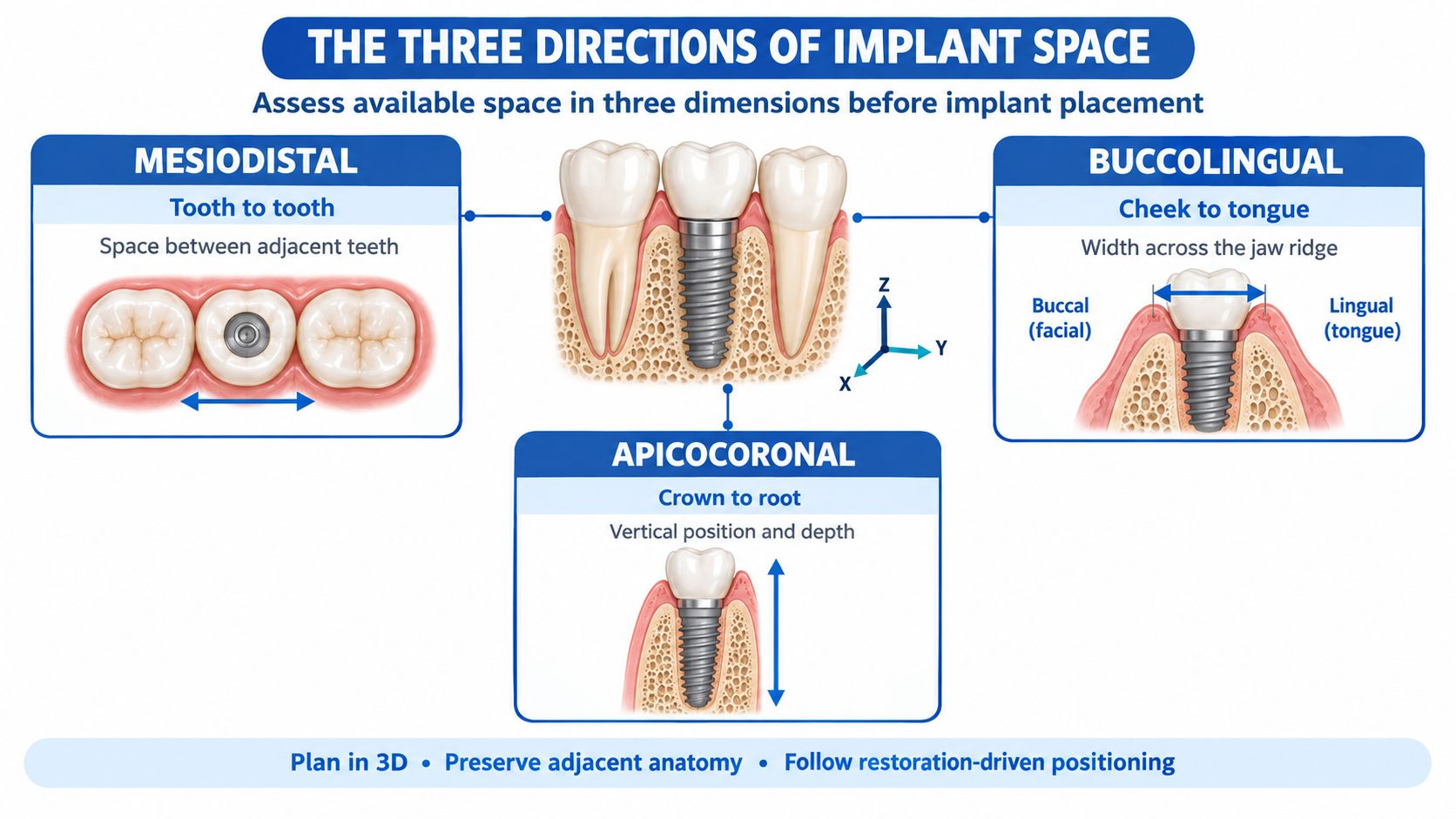
Mesiodistal space means side-to-side along the arch
Mesiodistal space is the room between teeth or between implants as you move along the curve of the jaw. It answers a simple question, can the implant sit without crowding a neighboring root or another implant? In the front and back of the mouth, that clearance helps protect the interproximal bone peak, the little bit of bone between teeth that supports the gum shape.
Buccolingual space means cheek-to-tongue width
Buccolingual space is the width of the ridge from the cheek side to the tongue side. That measurement matters because the implant has to fit inside the bone, not just within the visible gap. If the ridge is too thin, the facial bone and soft tissue can be at risk, especially where appearance matters most.
Vertical space means top-to-bottom restorative room
Vertical space is the height from the planned implant platform to the biting tooth opposite it. That room has to fit the abutment, the restorative material, and the crown or attachment. If the space is short, the restoration can become bulky, weak, or hard to clean.
A board-certified periodontist may use i-CAT 3D cone beam imaging to see all three directions at once. In a guided case, X-Nav dynamic surgical navigation can then help carry that plan into surgery, so the drill path follows the space that was measured rather than a guess made from a flat image. The language sounds technical, but the idea is plain, measure in three dimensions, then place the implant where those three dimensions work.
Mesiodistal Space for a Single Tooth Implant
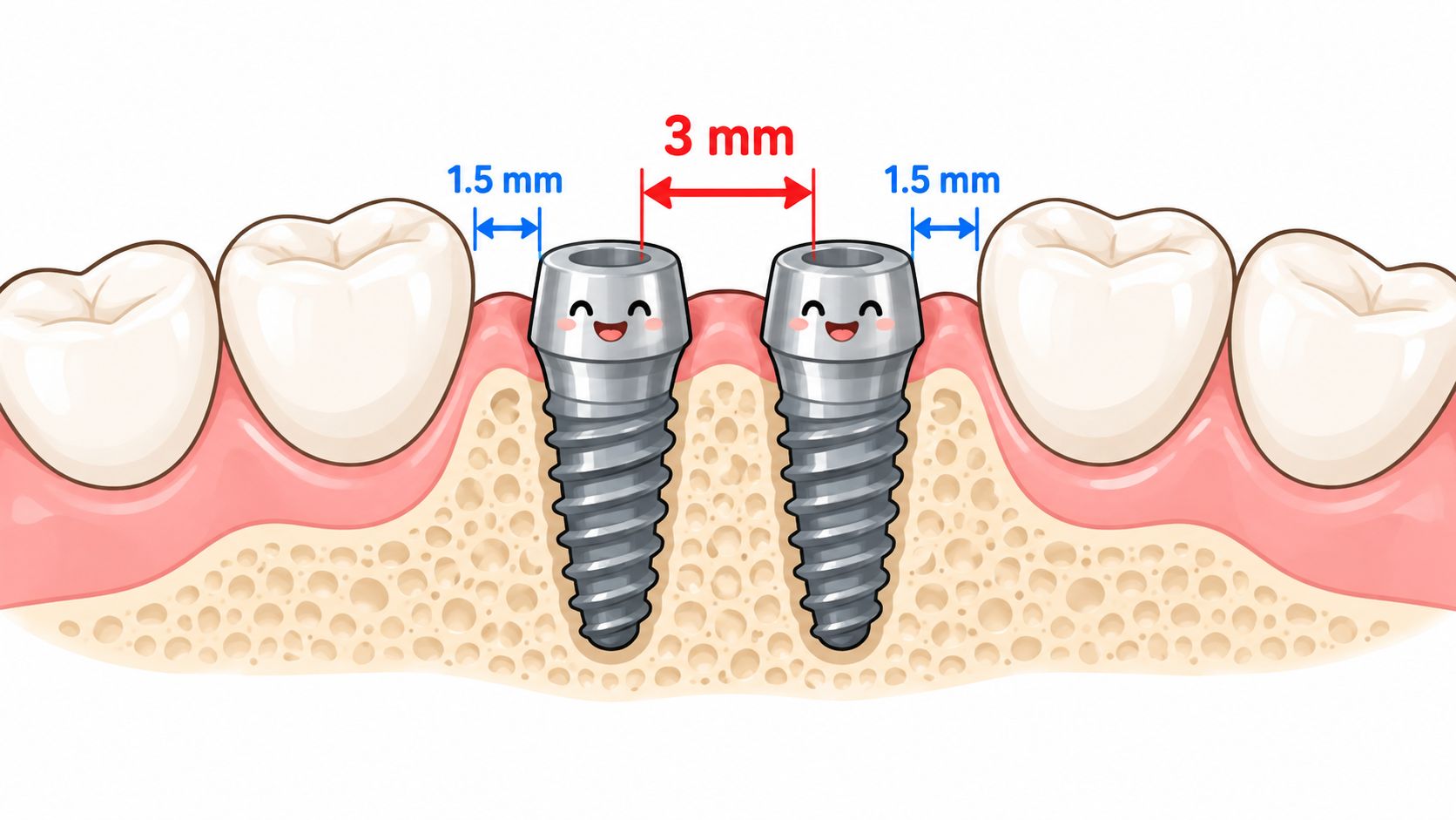
A single implant between two natural teeth usually needs about 7 mm of mesiodistal space when a standard 4 mm implant is used, because the implant itself takes up about 4 mm and each side needs about 1.5 mm of clearance to the adjacent root. That buffer is there to protect the neighboring tooth's periodontal ligament and the bone between the teeth. If the implant sits too close, the adjacent root and bone can pay the price.
Why the 1.5 mm buffer matters
That small gap is not an arbitrary cushion. It gives the bone and soft tissue enough room to stay stable around both the implant and the natural tooth. If placement gets closer than that, proximal bone remodeling can occur around the neighboring tooth, and bone height can drop.
What changes the total
The total number is not fixed for every mouth. A narrower implant may be considered in a tight anterior site, while a wider site may allow a different diameter or restorative shape. Root angulation matters too, because roots that lean toward each other can reduce usable room even when the crowns look separated.
A CBCT helps the clinician see whether the roots are straight, converging, or flared, and whether the final crown will land in a safe position. That's the part patients often don't see on a panoramic image. The visible opening can look generous, but the root anatomy underneath may tell a different story.
When a patient asks about treatment at Periodontal Associates of Delaware's Wilmington, Newark, or Dover offices, the answer usually starts with that scan-based check, not with a brand name for the implant. The numbers decide the starting point, and the anatomy decides whether a standard implant, a narrower implant, or another plan makes sense.
Interimplant Spacing When Two Implants Sit Next to Each Other
Two implants need their own buffer, and it's separate from the implant-to-root rule. The usual planning minimum is 3 mm between neighboring implants, measured between the implant shoulders or adjacent implant surfaces. That distance helps preserve the interimplant crestal bone and gives the soft tissue between the restorations a better chance to stay stable.
What happens when implants are too close
When the spacing falls below that benchmark, the zones of lateral bone remodeling can overlap. The result may be a flatter bone peak between the implants, which can make papilla fill less predictable. One clinical report summarized mean crestal bone-height loss of 1.04 mm when the 3 mm recommendation was violated, compared with 0.45 mm when the spacing was maintained (crestal bone spacing report).
That doesn't mean the soft tissue will always look poor if the spacing is tight, and it doesn't mean every wide spacing creates a perfect papilla. Bone level, tissue thickness, implant depth, and prosthesis design all play a role. The 3 mm rule is a planning benchmark, not a cosmetic guarantee.
Two adjacent implants need more than “enough room to fit.” They need room to preserve the bone peak between them.
What clinicians consider in tight multi-unit cases
When the site is narrow, the plan may shift toward a narrower implant, orthodontic space development, staged treatment, or a different prosthetic design. The choice depends on the final tooth positions, the bone volume, and how the restoration has to emerge through the gum. A patient hearing that explanation should take one message from it, the goal is not to force implants into a tight corridor. The goal is to choose the safest layout for the bone that's there.
Buccolingual Ridge Width and Facial Bone
From cheek to tongue, the ridge has to be wide enough to hold the implant and still leave bone where the soft tissue needs support. For a standard 3.7 to 4.0 mm implant, a commonly cited planning target is about 6 mm of ridge width, with roughly 1.5 to 2 mm of facial bone preferred for soft-tissue stability, especially in the esthetic zone (clinical guide on ridge width and facial bone). That facial side matters because it is the side patients see when they smile.
Why the anterior maxilla gets special attention
The upper front area is unforgiving when facial bone is thin. If the bone on the lip side is fragile, resorption can change the gum contour and make the final crown look longer or less natural. That's why ridge preservation after extraction can matter so much in the front of the mouth. It helps maintain the envelope that the future implant and crown need.
When grafting enters the conversation
If the ridge is too narrow, grafting may be discussed before implant placement. In the upper back jaw, the conversation can also involve sinus augmentation if the anatomy limits where the implant can sit. Those choices are about creating a stable foundation, not about adding complexity for its own sake.
For patients who want to understand bone rebuilding in more detail, Periodontal Associates of Delaware's explanation of periodontal bone grafts is a useful place to start. The key point is simple. Buccolingual space is not just width on a chart. It's the difference between an implant that sits inside a supported ridge and one that risks thin facial bone and a compromised emergence profile.
Vertical Restorative Space by Prosthesis Type
Vertical space is the part of implant planning patients most often hear about after the horizontal numbers are discussed. It's the distance from the planned implant platform, or ridge, up to the biting tooth opposite it. That space has to fit the attachment, the framework or acrylic, the tooth, and enough material thickness for strength and hygiene.
| Prosthesis Type | Approx. Minimum Vertical Space | Planning Notes |
|---|---|---|
| Fixed screw-retained restoration at implant level | 4 to 5 mm | Very compact, but design details still matter |
| Fixed screw-retained restoration at abutment level | 7.5 mm | Needs room for the abutment and restoration |
| Fixed cement-retained crown | 7 to 8 mm | Requires enough height for crown form and retention |
| Unsplinted overdenture | 7 mm | Compact option for selected removable cases |
| Bar overdenture | 11 mm | More vertical room is needed for the bar and prosthesis |
| Fixed screw-retained hybrid prosthesis | 15 mm | Requires the most vertical space among the listed designs |
That range shows why “more space” is not always better. Too little height can leave thin restorative material, poor retention, or a hard-to-clean emergence profile. Too much height can create long crowns and more twisting force on the implant, which can increase biomechanical loading.
Why attachment choice changes the plan
Ball attachments commonly need about 10 to 12 mm of vertical room, while more compact Locator or OT Equator-style attachments may need about 7 to 9 mm. A recent review also noted approximate minimums of 8.5 mm for one complete prosthetic setup and 7 to 7.5 mm for another compact setup (review of vertical restorative space). The exact attachment system changes the surgical and restorative plan, which is why the prosthesis needs to be decided before the implant goes in.
What the bite has to leave room for
A single crown can run short on space even when the bone is fine. Full-arch cases often need more room still. That's why a mounted cast or digital articulation matters. The periodontist and restorative team are checking not just whether an implant can fit, but whether the final restoration can be cleaned, tightened, and maintained without constant compromise.
How Tooth Position Changes the Numbers
Space requirements change by where the implant is going. A canine site doesn't behave like a first molar site, and a molar site doesn't behave like a front-tooth site. That's why a single universal number doesn't tell the whole story.
Different teeth, different room needs
A published spacing model proposed approximately 4 to 4.6 mm from an implant position to an adjacent canine, 7 to 7.4 mm for a subsequent position, 8 to 8.5 mm for a first-molar position, and 9 to 9.5 mm for a second-molar position. It also reported about 4 to 4.6 mm adjacent-to-implant spacing for premolars and 5 to 5.5 mm for molars (tooth-position spacing study).
Those differences make sense. Posterior teeth need more room because the crowns are larger and chewing forces are higher. Anterior teeth are driven more by esthetics, gum shape, and papilla support.
Why this matters during planning
A patient can hear “there's enough room” and still not have enough room for the tooth position that's being restored. A molar implant may demand a different layout than a premolar implant in the same mouth. That's why a clinician will check the final crown position, not just the empty gap.
In a Delaware periodontal consult, that often means the scan gets read alongside the proposed tooth number, not in isolation. The implant is being placed for a specific crown, in a specific part of the arch, with specific biting forces. That detail is what turns a raw measurement into a usable treatment plan.
Putting the Numbers Together With CBCT and Navigation
A CBCT scan shows the site in three dimensions, which matters because the mouth doesn't offer a flat problem. It shows root angulation, ridge width, facial bone thickness, sinus position, and undercuts that can change implant diameter choice. It also shows whether the planned crown position lines up with the bone, which is the heart of prosthetically driven planning.
What the scan adds that a flat image can't
A panoramic image can give a broad overview, but it can hide convergence, thin facial bone, or a ridge that narrows quickly inside the gum. A CBCT helps the clinician understand whether the implant needs to move slightly, whether a narrower implant makes sense, or whether grafting should be part of the sequence. In other words, it's not a pass-fail check.
A CBCT is a decision tool, not just a yes-or-no screen.
How the surgical plan gets carried out
Once the plan is built, dynamic navigation such as X-Nav can guide the drill path during surgery. That matters when the margins are tight, because a few millimeters in the wrong direction can change the outcome. The goal is to place the implant where the restorative plan, bone volume, and soft tissue all agree.
For some Delaware patients, that scan-based process may also be paired with periodontal therapy first if inflammation or bone loss is part of the picture. Periodontal Associates of Delaware uses advanced imaging and implant planning to connect the restorative goal with the surgical path, so the scan, the bite, and the final tooth all match the same plan.
Options When the Space Is Too Tight
A narrow site does not automatically rule out an implant. It usually changes the sequence of care. The answer may be to move teeth, preserve or rebuild bone, or choose a different restoration so the final plan fits the available room.
Common ways clinicians may create room
- Narrower implants: A slimmer implant can sometimes fit where a standard diameter cannot.
- Orthodontic space opening: Teeth can sometimes be moved to create safer mesiodistal room.
- Extraction-site preservation: Ridge dimensions can be maintained after tooth removal so the site does not collapse further.
- Sinus lift and implant at the same time: Bone can be rebuilt when width or height is lacking, including selected posterior cases.
- Alveoloplasty: Selected bone reshaping can create better restorative room in the right case.
- Occlusal equilibration: Small bite adjustments may help create needed vertical clearance.
- Orthodontic intrusion: A supraerupted tooth may be moved back to regain vertical room.
- Prosthetic redesign: A different attachment or abutment-level versus implant-level restoration may fit the anatomy better.
If active gum disease is part of the picture, periodontal treatment may need to happen first. In that setting, LANAP with the FDA-cleared PerioLase MVP-7 may be one of the treatment paths considered, depending on the case. The sequence has to match the problem.
A tight space is often a three-dimensional problem. One patient may need more width, another more height, and another a small change in tooth position before the implant can be placed safely. A Delaware periodontal office may sort that out on CBCT, then decide whether movement, grafting, or a different prosthetic design gives the best path forward.
When the plan changes instead of the space
Sometimes the responsible choice is to rebuild bone, stabilize gum health, or redesign the restoration rather than force an implant into the smallest available gap. Patients often feel more settled when they hear that a limited site can still be treated, just not always with the fastest or simplest sequence.
Planning Minimums Versus Patient-Specific Guarantees
The numbers in this article are planning benchmarks, not promises. Two patients can have the same measured gap and still need very different plans because implant diameter, tissue thickness, opposing teeth, bite forces, esthetic demands, and grafting needs all change the decision. A thin gum biotype, for example, behaves differently from a thick one. A heavy grinder also asks for a different plan than a light chewer.
A board-certified periodontist uses those same minimums, but the final target is patient-specific. That's where consultation matters, because the scan is only one piece of the picture. The soft tissue, the bite, the future crown, and the maintenance plan all have to fit together.
For readers who want a deeper look at how bone and gum support are evaluated, guided tissue regeneration is one of the periodontal concepts that can come into play when rebuilding support is part of the treatment plan. Individual results vary, and the safest next step is a consultation with a periodontist who can read the scan in the context of your own anatomy.
Quick-Reference Numbers and Next Steps
Here's a compact way to remember the main benchmarks. 1.5 mm between an implant and a natural root, 3 mm between two implants, about 7 mm for a standard single-tooth site, about 6 mm of buccolingual width, 1.5 to 2 mm of facial bone, and a vertical height that depends on the prosthesis (root and implant spacing guidance). That's the language a periodontist is using when the CBCT is on the screen.
| What to Ask About | The Working Number | Why It Matters |
|---|---|---|
| Implant to adjacent root | 1.5 mm | Protects the root and nearby bone |
| Implant to implant | 3 mm | Helps preserve the bone peak between implants |
| Single-tooth mesiodistal site | About 7 mm | Fits a standard implant with clearance |
| Buccolingual ridge width | About 6 mm | Supports a standard implant body |
| Facial bone | 1.5 to 2 mm | Supports soft-tissue stability |
| Vertical space | Depends on prosthesis | Must fit the chosen crown or attachment |
If the numbers sound tight, that doesn't automatically rule out treatment. It just means the plan has to be built carefully, in the right sequence, with the right imaging.
Periodontal Associates of Delaware helps patients evaluate implant space, bone volume, gum health, and the bite before treatment moves forward. If you're trying to understand whether there's enough room for an implant, visit Periodontal Associates of Delaware and request an appointment at the nearest Wilmington, Newark, or Dover office so a periodontist can review your scan and discuss your options.