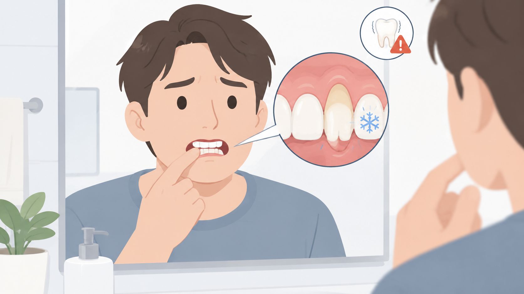
A tooth looks longer in the bathroom mirror than it did last year. Cold water suddenly stings near the gumline, and a small yellowish area appears where pink tissue used to sit. It's understandable to worry about tooth loss or imagine that gum surgery is inevitable.
For adults searching for information about teeth, gums receding, and exposed roots, the most useful starting point is the cause. The same exposed root can result from inflammation, brushing trauma, thin tissue, frenum pull, or bite-related problems. Those differences affect both the treatment needed and how much coverage may be realistic.
Table of Contents
- What Longer Teeth Usually Mean
- Why Gums Recede Beyond Hard Brushing
- Sensitivity, Bare Roots, and Loose Teeth
- Everyday Care That Protects the Gumline
- How 3D Imaging Shapes the Plan
- Grafts, Frenectomy, and Restorative Choices
- Book a Recession Evaluation Near You
What Longer Teeth Usually Mean
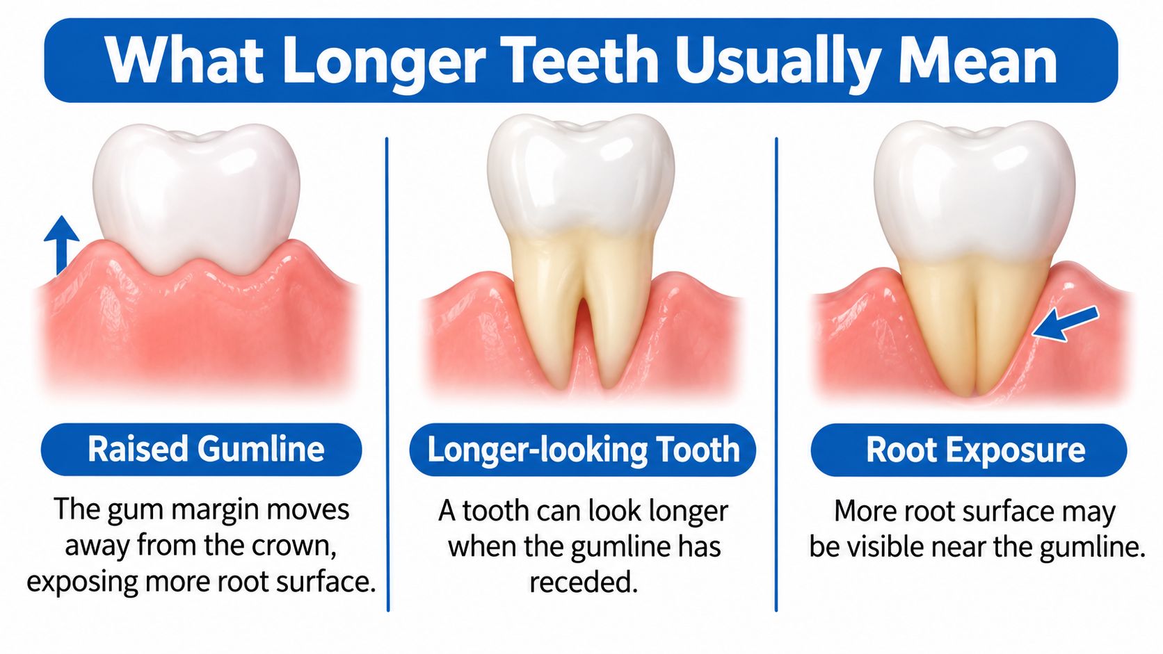
A longer-looking tooth hasn't necessarily moved or grown. Gingival recession means the gum margin has moved away from the crown, exposing part of the root. A person might notice this beside a canine while the neighboring teeth still look unchanged.
The exposed area may look darker than the crown or feel like a small step near the gumline. Appearance alone, however, can't establish whether the site is stable or whether supporting tissues are being damaged.
Measuring the change rather than guessing
A periodontist measures recession from the cementoenamel junction, where crown enamel meets the root, to the free gum edge. A calibrated periodontal probe provides a repeatable measurement rather than a visual estimate.
A standardized full-mouth examination records six sites per tooth across 28 teeth, excluding third molars, because recession can differ sharply between neighboring surfaces, as described in the clinical measurement summary indexed by PubMed. This creates a baseline for comparing future visits.
Recession prevalence in U.S. adults, 2009 to 2014
An analysis of the National Health and Nutrition Examination Survey evaluated 10,676 participants, representing approximately 143.8 million U.S. adults, and reported the following findings using a threshold of at least 1 millimeter of mid-buccal recession at one or more sites.
| Measure | Adults affected |
|---|---|
| Recession of 1 mm or more, whole mouth | 91.6% |
| Recession of 1 mm or more, aesthetic zone only | 70.7% |
| RT1 recession, whole mouth | 12.4% |
| RT1 recession, aesthetic zone | 5.8% |
Mid-buccal means the middle of the cheek-facing tooth surface. The aesthetic zone refers to the teeth assessed within the visible smile area, while RT1 is a clinical recession classification, not a rating of pain or urgency.
Common doesn't mean every affected site needs surgery. A small finding may be stable and symptom-free; its location, progression, and surrounding tissue health matter.
For adults in Wilmington, Newark, Dover, and nearby New Castle and Kent County communities, a longer-looking tooth is a reason to identify the cause rather than assume the worst. Individual findings and results vary, so a periodontist should interpret the chart in the context of the whole mouth.
Why Gums Recede Beyond Hard Brushing
A stiff toothbrush often gets the blame, but brushing isn't the only explanation. A 2025 systematic review of recession risk factors reported the associations below, expressed as odds ratios rather than the probability that an individual will develop recession.
Why an exposed root can have different causes
| Factor | Reported odds | Pathway | What a clinician looks for |
|---|---|---|---|
| Periodontitis | 9.90 | Inflammatory | Bleeding, pockets, bone loss |
| High frenum attachment | 4.58 | Anatomical | Tissue pulled when the lip moves |
| Dental plaque | 4.26 | Inflammatory | Soft deposits at the gumline |
| Occlusal trauma | 3.20 | Bite force | Heavy contacts or a shifted tooth |
| Alcohol consumption | 2.04 | Lifestyle | Intake history with other risks |
| Prior periodontal treatment | 1.86 | Association, not proof of cause | Past care plus current chart |
| Smoking | 1.84 | Lifestyle | Tobacco use and slower healing |
| Hard or abrasive brushing | No figure reported in this review | Mechanical | Notches on cheek-facing roots |
These figures describe associations across studied groups. They aren't a diagnostic checklist, and they don't prove that a particular factor caused someone's exposed root.
Different paths can leave a similar appearance
Periodontitis damages the tissues that support teeth. Recession accompanied by bleeding, deep pockets, and bone loss raises a different concern from an isolated, uninflamed area of root exposure.
A high frenum attachment involves the band of tissue connecting the lip or cheek to the gums. If that attachment pulls close to the margin, lip movement may place tension on already vulnerable tissue.
Dental plaque collects around tooth surfaces and contributes to inflammation. A person can brush regularly yet miss deposits between teeth or along an irregular gumline.
Occlusal trauma refers to injury associated with excessive biting forces. Heavy contacts, tooth position, and clenching deserve assessment, but grinding shouldn't automatically be declared the cause of recession. The practice's explanation of grinding and receding gums explores that distinction.
Alcohol intake and smoking belong in the health history because they may accompany other risks. Smoking also matters when planning tissue healing.
An exposed root identifies a change in gum position. It doesn't identify the cause.
Why the history matters
A history of periodontal treatment is an association, not proof that treatment caused recession. Previous disease, changes after inflammation subsides, and the current condition all need consideration.
Brushing trauma may appear as wear or a notch on cheek-facing roots, particularly around canines and premolars. Still, a notch alone doesn't establish the cause. Thin tissue and a tooth positioned toward the edge of its supporting bone may increase vulnerability even when cleaning habits are careful.
During an evaluation, the periodontist checks probing depths, bleeding, tissue thickness, frenum pull, tooth position, and bite contacts. The important question isn't whether the root can be hidden. It's whether the tooth's support is healthy and whether the process causing the recession remains active.
Sensitivity, Bare Roots, and Loose Teeth
Receding gums don't automatically mean a tooth will fall out. A sensitive root and a tooth losing supporting bone can look similar in the mirror, yet they need different care.
Exposed dentin contains tiny channels that can transmit temperature and other stimuli toward the nerve. Unlike the crown, the root doesn't have a protective enamel covering, so cold drinks, sweets, or air may cause a brief zing.
The difference between recession and a pocket
Recession measures exposed root height. Pocket depth measures the space between the gum edge and the tooth, so the measurements answer different questions.
The National Institute of Dental and Craniofacial Research's gum disease guide explains that healthy periodontal pockets are usually 1–3 millimeters deep, while deeper pockets may indicate periodontal disease; dental X-rays can reveal bone loss, and advanced loss of supporting bone can allow teeth to loosen or shift.
A periodontist interprets pocket depth together with bleeding, attachment levels, and imaging. A shallow, uninflamed site with visible root exposure is different from a deep, bleeding pocket beside reduced bone support.
Symptoms and what they may signal
| Sign | Usually points to | Next step |
|---|---|---|
| Short zing to cold, sweet, or air with a clean, shallow gumline | Exposed dentin sensitivity | Desensitizing care and monitoring |
| Bleeding gums with pockets deeper than the healthy range above | Periodontal inflammation or disease | Periodontal charting and cleaning |
| Tooth looks long and feels slightly loose | Possible bone loss | Probing plus dental X-ray |
| Food trapping between teeth, with teeth seeming to shift | Possible attachment or bone loss | Individualized evaluation |
Changes that deserve prompt attention
A growing defect, inadequate tissue thickness, or new mobility deserves timely assessment. A person shouldn't keep testing a loose tooth with fingers or the tongue, because repeated movement won't clarify the diagnosis.
The practice's guide to tooth mobility explains why movement needs an individualized evaluation. Mobility can have more than one contributing factor, so treatment shouldn't be chosen from appearance alone.
Practical distinction: Sensitivity describes what a tooth feels. Bone and attachment measurements help explain how well it's supported.
Identifying damage before more support is lost may allow a stronger focus on preserving the tooth. Persistent or lingering pain also warrants assessment because not every painful tooth has a recession-related problem. Bleeding, sensitivity, shifting, and apparent lengthening should be charted, not diagnosed from a photograph.
Everyday Care That Protects the Gumline
Use a soft brush with light pressure. Small, controlled circles or a gentle electric-brush technique can clean the gumline without repeatedly scrubbing the exposed root. Bristles should contact the margin without flattening against the tooth.
Clean between teeth daily. Floss works well for tight contacts; an interdental brush may suit wider spaces. The brush should fit comfortably rather than require force, and a dental professional can demonstrate the appropriate size.
Use sensitivity toothpaste consistently. Follow the product directions and allow days to weeks rather than judging the result after a single brushing. Persistent symptoms need examination instead of repeated product changes.
Stop tobacco use. Smoking is associated with recession and can interfere with healing. Cessation support can help make this change more manageable.
Report alcohol use, grinding, and clenching. A complete history helps the clinician consider lifestyle risks and bite-related stress without assuming that every exposed root has the same origin.
Keep periodontal maintenance appointments. Once periodontal disease is present, professional follow-up helps assess areas that can't be evaluated or cleaned adequately at home. Waiting for pain can delay recognition of a problem.
Thin tissue needs a tailored approach
Some people have naturally thin, scalloped gums rather than a broad band of thicker tissue. A prospective orthodontic cohort summarized in a review of recession and tissue-related risk identified a thin-scalloped phenotype as the strongest predictor of new recession, with odds approximately 4.1 times higher than in comparison phenotypes.
That finding doesn't mean thin gums will recede. It means tissue anatomy deserves attention when choosing cleaning methods, planning tooth movement, or deciding whether added tissue thickness may be useful.
Progress should be measured, not assumed
Less bleeding and easier cleaning are useful changes, but they don't establish that every site is stable. A professional comparison of gum measurements provides a clearer answer than checking the mirror each morning.
Many patients may notice less sensitivity after gentler cleaning and desensitizing care, even while some root remains visible. If discomfort persists or the exposed area grows, professional treatment may still be needed.
Home care can reduce plaque, inflammation, and avoidable trauma, but it won't pull a receded gumline back over a root or release a tight frenum.
How 3D Imaging Shapes the Plan
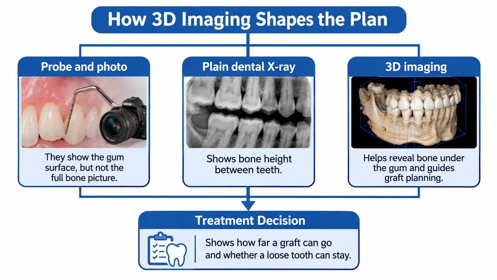
A probe measures the gum-tooth relationship, and a photograph records surface appearance. Neither provides the complete picture of the supporting bone.
A conventional dental X-ray helps show bone height between teeth. When additional information would change treatment, cone-beam imaging can add views of ridge width, shape, and the relationship between a root and its bony housing.
The curtain and the stage
Gum tissue is like a curtain, while supporting bone is the stage beneath it. Moving the curtain can change coverage, but it doesn't rebuild a stage that has already shrunk.
This distinction helps explain why a graft may improve tissue thickness without completely restoring the former gumline. Bone levels between teeth, root position, and neighboring tissue all influence the plan; imaging supports that assessment rather than replacing the examination.
Decisions imaging may change
- Separate tissue position from structural loss. Images can help clarify whether the concern involves gum position alone or a broader loss of support. A cone-beam scan isn't necessary for every recession site and may not reliably display extremely thin bone.
- Plan replacement only when needed. If a tooth can't reasonably be retained, the clinician can evaluate an implant path and whether ridge grafting or sinus augmentation may be needed before placement.
- Coordinate surgical and restorative boundaries. Imaging, examination, and restorative planning help determine where sound tooth structure remains for a restoration or crown-lengthening procedure.
Periodontal Associates of Delaware uses i-CAT 3D cone beam imaging, X-Nav dynamic surgical navigation for implants, and the FDA-cleared PerioLase MVP-7 for LANAP. Dynamic navigation can guide implant placement in real time using the scan-based plan, but no technology removes every surgical risk.
A scan is a planning step, not a commitment to surgery. Imaging should be selected for a clinical reason, with the expected benefit weighed against radiation exposure.
Patients may review the findings with the periodontist and general dentist before choosing monitoring, disease treatment, grafting, or replacement. Individual anatomy varies, and an image helps define realistic choices rather than guarantee an outcome.
Grafts, Frenectomy, and Restorative Choices
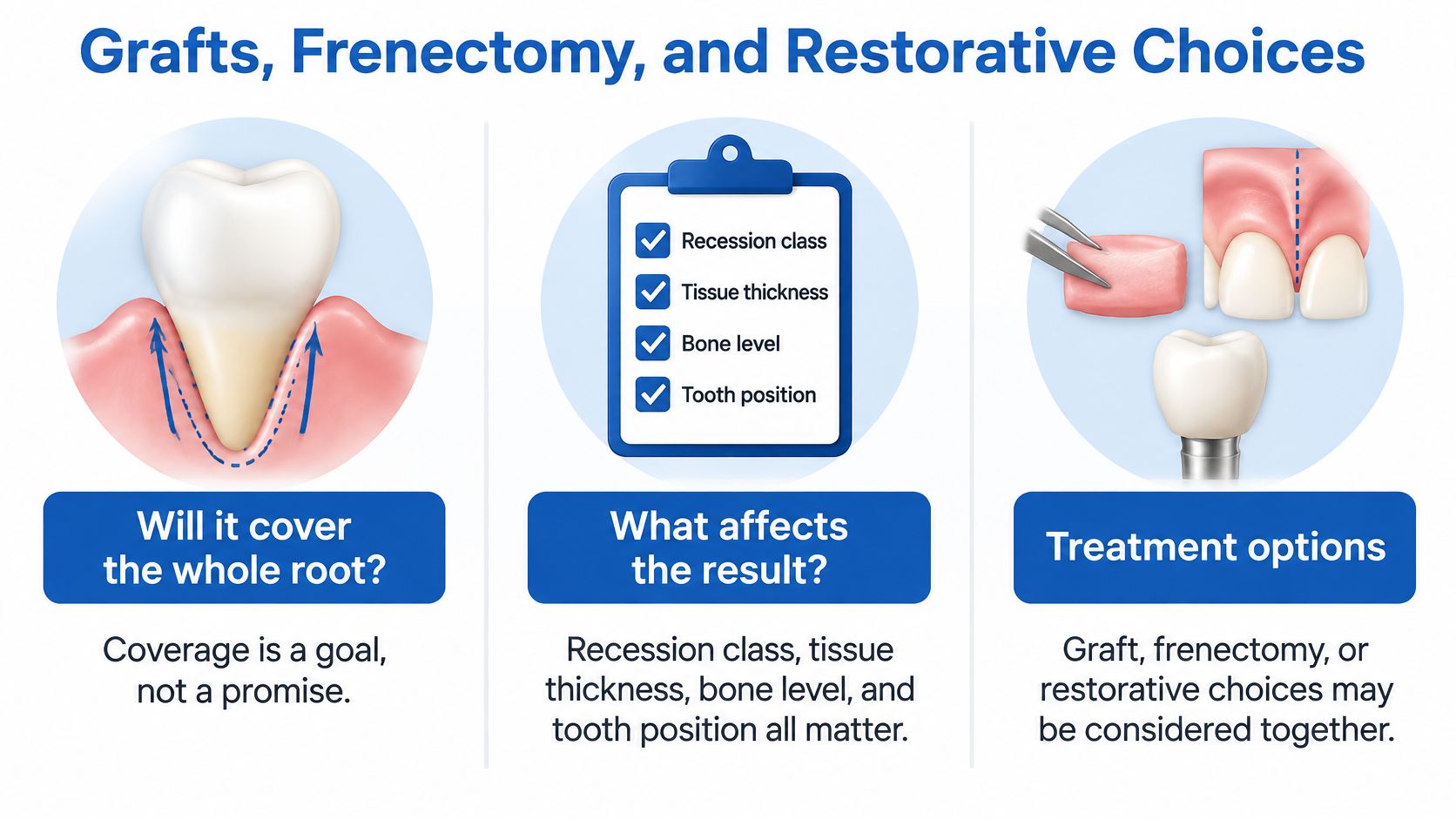
Will a graft cover the whole root? Coverage is a goal, not a promise. Recession classification, tissue thickness, bone levels, tooth position, and root wear all affect what's achievable.
A treatment plan should name its main objective. Reducing sensitivity, adding durable tissue, improving appearance, and achieving complete coverage aren't identical outcomes.
Clinical options when gums have receded
| Option | Main job | What changes | Often considered when |
|---|---|---|---|
| Connective-tissue graft | Root coverage and thicker gum | Palate tissue placed over the root | Sensitivity, thin gum, or esthetic root show |
| Acellular dermal matrix | Coverage without a large palate harvest | Donor substitute under the gum | Several sites or limited palate tissue |
| Frenectomy | Stop a tug on the margin | Releases a high frenum | Lip or cheek pull linked to the defect |
| Crown lengthening | Expose tooth for a restoration or even the gumline | Removes gum and sometimes bone | A crown needs more tooth, not root coverage |
| Root filling or bonding | Repair a worn notch | Rebuilds tooth, not gum position | A cervical lesion sits on the bare root |
| LANAP laser therapy | Treat periodontal infection | Targets diseased pockets with PerioLase MVP-7 | Recession occurs alongside periodontal disease |
Graft materials involve trade-offs
A connective-tissue graft commonly uses tissue from the palate, combined with movement of the existing gum toward the crown. The practice's explanation of a subepithelial connective-tissue graft describes this approach.
A 2025 systematic review comparing graft materials found that 11 of 14 randomized trials judged acellular dermal matrix as effective as connective-tissue grafts for Class I and II recession, while three trials found better coverage with connective-tissue grafts. Substitute materials may be useful when treating several sites would otherwise require substantial palate tissue.
Donor-site morbidity means symptoms or complications where tissue is collected, such as palatal soreness or bleeding. Avoiding a large donor site may be valuable, but material choice still depends on the defect and treatment objective.
A systematic review of coronally advanced flap procedures found that adding connective tissue or enamel matrix derivative improved the likelihood of complete root coverage compared with the flap alone. That doesn't make complete coverage predictable for every root.
Each procedure has a different job
A frenectomy releases a pulling tissue attachment. It doesn't cover an exposed root by itself. Crown lengthening serves almost the opposite purpose of root coverage: it exposes more tooth for restorative access or selected smile corrections.
When a worn notch accompanies recession, bonding or a filling repairs tooth structure while periodontal surgery addresses tissue position and thickness. A systematic review of recession with non-carious cervical lesions supports coordinated planning, while noting differences among studies that limit firm conclusions.
A useful treatment goal: A more stable, less sensitive site can be worthwhile even when some root remains exposed.
Disease control and recovery come first
Active periodontal infection needs treatment before elective root coverage. LANAP may be considered for appropriate periodontal disease sites; it isn't a substitute for a root-coverage graft.
Many patients may experience less pain and a quicker return to routine activity with LANAP than with traditional flap surgery. It isn't appropriate for every defect, and neither laser treatment nor grafting should be presented as pain-free.
Individual results vary. Before choosing treatment, anxious patients can request an explanation of the procedure, discomfort management, expected healing, donor-site care, and what partial coverage might look like at the specific site.
Book a Recession Evaluation Near You
A tooth may look longer for different reasons, and the same exposed root can reflect gum disease, a pulling frenum, thin tissue, bite force, or brushing trauma. The cause matters: it shapes what treatment can realistically cover. An evaluation can establish baseline measurements, even when the area is not painful.
If you have concerns beyond the symptoms described in the earlier sensitivity section, consider booking an assessment when root exposure appears to be increasing or food repeatedly catches in a widening space.
What to expect from the appointment
The periodontist reviews your health history and measures the gums around your teeth. They may also assess tissue thickness, frenum pull, tooth position, and bite. Imaging may help when bone loss, structural concerns, or implant planning could affect the treatment decision.
Bring a list of questions, along with any changes you have noticed since your last dental visit. Ask which cause seems most likely, whether monitoring is reasonable, what each suitable treatment involves, how healing may affect your routine, and what partial root coverage could mean for that tooth.
Access across Delaware
The periodontists at Periodontal Associates of Delaware hold diplomate status with the American Board of Periodontology. The practice has locations in Wilmington, Newark, Dover, and Rehoboth Beach, and serves patients in Pike Creek and Hockessin.
A referral isn't needed to schedule an appointment. Most major insurance plans are accepted, and flexible financing is available. Insurance acceptance does not establish coverage for a particular procedure.
Periodontal Associates of Delaware provides evaluation and care for gum recession, exposed roots, and the tissues supporting teeth. To discuss monitoring, grafting, or periodontal disease treatment, visit Periodontal Associates of Delaware and request an appointment at a convenient office.