A tongue catches a small notch behind a tooth. Cold water suddenly stings near the gumline, or food keeps collecting in a space that didn't seem noticeable before. For an adult already worried about gum surgery, that discovery can feel unsettling. The most useful next step isn't guessing whether a graft is needed. It's finding out exactly where the gum has changed and what's happening beneath it.
Table of Contents
- Noticing a Receding Gum Behind a Tooth
- What Recession Actually Means on the Back of a Tooth
- Why the Back of a Tooth Recedes in the First Place
- Do You Need Treatment or Just Monitoring
- What Happens at a Periodontal Evaluation
- Modern Options for Treating Posterior Recession
- Recovery and What Most Patients Actually Experience
- Key Takeaways and Next Steps for Delaware Patients
Noticing a Receding Gum Behind a Tooth
A search for “receding gum back of tooth” can describe different concerns. Some adults mean the tongue-facing surface of a lower tooth. Others mean the palate-facing surface of an upper tooth or the gum around a molar at the back of the mouth. Those locations aren't interchangeable, so identifying the exact surface helps the clinician narrow the examination.
The first clue may be a longer-looking tooth, a newly visible root, or sensitivity during brushing. Food trapping can draw attention to the area, too, although that symptom alone doesn't establish recession. The clinical term gingival recession describes a gum margin that has moved away from the tooth's crown, exposing part of its root.
A common finding, not a diagnosis of severe disease
Recession is widespread, but frequency shouldn't be confused with severity. A systematic review reported a pooled prevalence of 84.92% when recession was defined as at least 1 millimeter, while emphasizing that estimates change with the population, examination method, tooth location, and measurement threshold (systematic review of gingival recession prevalence).
That distinction matters for an anxious patient. A small exposed area doesn't, by itself, establish advanced gum disease or predict tooth loss.
- A visible change needs context. Its appearance doesn't reveal whether the tissue is stable or actively changing.
- Symptoms help guide the visit. Cold sensitivity, bleeding, or a change in tooth position deserves mention.
- An examination provides a baseline. Recorded measurements allow later comparisons instead of relying on memory or mirror checks.
Practical starting point: A new-looking gumline is a reason for an assessment, not proof that surgery is inevitable.
A useful consultation begins with the patient's observations: where the change appeared, whether it hurts, and whether earlier dental photographs or records show the same area.
What Recession Actually Means on the Back of a Tooth
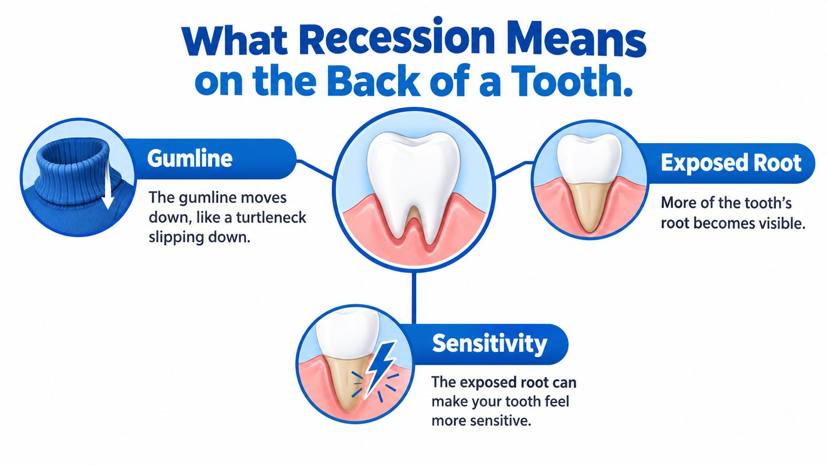
The gumline can be compared with a turtleneck collar around a tooth. When the collar shifts toward the root, it exposes a surface that was previously covered. On upper teeth, that movement may look upward; on lower teeth, it may look downward. Recession means movement toward the root tip, not movement in one visual direction.
The crown is covered by enamel. An exposed root has different surface protection, and exposed root dentin is more vulnerable to sensitivity, abrasion, plaque retention, and cervical decay. Clinically, recession is an apical displacement of the gum margin with root exposure (clinical discussion of recession mechanisms and assessment).
The visible edge isn't the whole support system
A tooth's support includes gum attachment and the surrounding bone. The exposed root is the part visible in a mirror, but that view doesn't show the condition of the supporting tissues between teeth or beneath the gumline.
Two separate findings therefore matter:
- Recession describes the position of the gum margin.
- Attachment loss and pockets describe other aspects of the tooth's supporting tissues.
A shallow exposed root may have a different treatment pathway from an area with inflammation and loss of support. Neither the size of a photograph nor the apparent length of a tooth can settle that distinction.
For a tongue-facing defect, visibility and access can make inspection and cleaning more difficult. Around a back tooth, the clinician also needs to evaluate the particular root shape, tissue thickness, and surrounding anatomy rather than assume that every posterior site has thin gums.
Location changes what must be examined, not just how the defect looks. A cheek-facing illustration can explain recession, but it can't substitute for examining the actual tongue-side or posterior surface.
Why the Back of a Tooth Recedes in the First Place
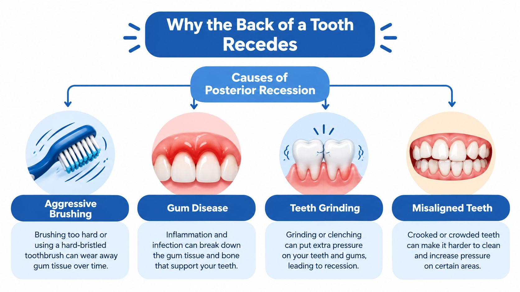
A notch behind a tooth shouldn't automatically be blamed on hard brushing. Brushing trauma is one possibility, but local anatomy, tooth position, periodontal inflammation, and bite-related factors can overlap. A systematic review found associations with plaque, high frenum attachment, occlusal trauma, and periodontitis, supporting a broader assessment rather than a single-cause explanation (systematic review of factors associated with recession).
Several contributors may act together
- Brushing technique and tissue resistance. Repeated scrubbing can traumatize the gumline, particularly where protective tissue is limited. The question isn't merely whether a patient brushes often, but how the brush contacts that particular surface.
- Tooth position and orthodontic history. A root positioned toward or outside its supporting bony housing may have limited tissue protection. Previous tooth movement belongs in the history, but orthodontic treatment alone doesn't prove the cause.
- Frenum attachment. A frenum is a fold of soft tissue. Its attachment can matter when evaluating local tissue tension and the amount of protective gum.
- Plaque and restorative contours. A hard-to-clean lingual surface or an overhanging restoration may contribute to plaque retention and inflammation.
- Bite forces and tobacco exposure. Grinding, bite contacts, and smoking warrant discussion, without assuming that any single factor explains the defect.
The relationship between teeth grinding and receding gums requires that same caution. A history of clenching doesn't replace an assessment of inflammation, tooth position, and support.
Similar appearances can require different decisions
Consider a hypothetical comparison. An isolated exposed root with thin, clean tissue and unchanged measurements may call for protection and observation. A similarly sized exposed area with bleeding and a deep pocket raises a different concern.
These aren't interchangeable diagnoses. The first situation centers on tissue vulnerability; the second requires investigation of active disease. The location and measurements help distinguish them, while the patient's habits and history help explain why the problem developed.
Do You Need Treatment or Just Monitoring
Not every recession defect needs grafting. A periodontist first identifies the problem treatment is meant to solve: progression, inflammation, sensitivity, root damage, or inadequate protective tissue. The visible amount of recession is only part of that decision.
Lost gum tissue doesn't regrow after a change in brushing technique. However, controlling the cause may help prevent worsening, and an exposed root may be managed with monitoring, sensitivity treatment, periodontal therapy, or grafting depending on the examination (patient guidance on recession and treatment selection).
Matching Recession Signs to Likely Next Steps
| What You Notice | Likely Category | Typical Next Step |
|---|---|---|
| A shallow area that appears unchanged and causes no symptoms | Possibly stable recession | Establish measurements, preventive care, and monitoring |
| Cold sensitivity without obvious swelling | Exposed-root symptoms | Assess the root and discuss desensitizing care |
| Bleeding or persistent inflammation | Possible periodontal disease | Evaluate pockets and control inflammation |
| A notch, darkened root area, or food-trapping contour | Possible wear, decay, or restorative issue | Coordinate periodontal and restorative assessment |
| Increasing recession or inadequate protective tissue | Possible need for tissue augmentation | Discuss whether grafting fits the anatomy and goals |
| Pus, mobility, or changing tooth position | Possible active supporting-tissue problem | Arrange prompt periodontal assessment |
The table describes reasons for evaluation, not diagnoses. A root notch, for example, might require restorative planning rather than a graft alone. Sensitivity also needs assessment instead of being attributed automatically to recession.
The useful question isn't only whether the gum can cover the root again. It's whether the immediate goal is stability, comfort, disease control, or coverage.
Monitoring should be an active plan with documented findings and professional review. It shouldn't mean ignoring a change until pain develops. Conversely, an asymptomatic, stable defect shouldn't be treated as an automatic surgical emergency.
When active periodontal inflammation is present, controlling that disease comes before an elective root-coverage plan. That sequence allows treatment decisions to reflect the underlying condition rather than appearance alone.
What Happens at a Periodontal Evaluation
A periodontal visit is an investigation, not a commitment to surgery. The clinician asks about the change, examines the tooth and surrounding tissues, and explains which findings affect the available choices. Patients can also describe their concerns about discomfort, recovery, or previous dental experiences.
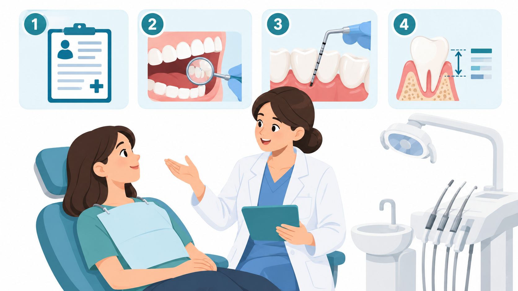
What the clinician checks
- Medical and dental history. Relevant details include medications, tobacco use, orthodontic treatment, brushing habits, restorations, and the timing of symptoms.
- The recession itself. The clinician measures its depth and width, examines root-surface changes, and evaluates tissue thickness and the band of protective keratinized gum.
- Attachment and inflammation. A periodontal probe helps assess pocket depth, bleeding, and attachment around the tooth. The distinction between recession and pocketing around teeth is important because an exposed root doesn't reveal pocket depth.
- Supporting anatomy. Tooth position, frenum attachment, bite contacts, and bone support help determine whether the proposed treatment fits the defect.
Photos can document appearance, but measurements and appropriate imaging answer different questions. The clinician selects imaging according to the clinical concern rather than assuming that every recession defect needs a three-dimensional scan.
Technology when it answers a specific question
Across the Wilmington, Newark, and Dover offices, the practice offers i-CAT 3D cone beam imaging when indicated for anatomical assessment and treatment planning. If a separate missing-tooth problem requires implant planning, X-Nav dynamic surgical navigation is available for that purpose; it isn't a treatment for an exposed root.
A LANAP-certified clinician can also assess whether laser-assisted periodontal treatment fits coexisting gum disease. The consultation should distinguish that disease-control discussion from the separate question of root coverage.
Appointment length depends on the examination and imaging needed, so the scheduling team can clarify the expected visit. Before recommending treatment, the periodontist should explain what was found, what remains uncertain, and which goals are realistic.
Modern Options for Treating Posterior Recession
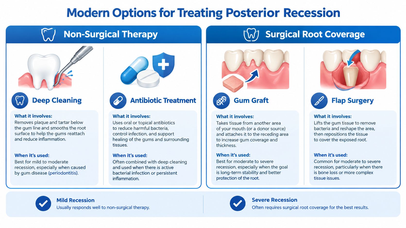
Treatment selection depends on the defect, supporting tissues, and intended goal, not just whether recession looks mild or severe. Disease-control therapy and root-coverage surgery address different problems, and some patients need coordinated care.
Disease control and root protection
Non-surgical periodontal therapy, including scaling and root planing, may be recommended for periodontal disease. Desensitizing products, changes in traumatic habits, and correction of plaque-retaining restoration contours may address other parts of the problem.
For appropriate periodontal disease cases, the practice offers LANAP laser gum surgery using the FDA-cleared PerioLase MVP-7. This minimally invasive option belongs in the infection and periodontal-treatment discussion. It shouldn't be presented as interchangeable with a graft or as a promise to cover a recessed root.
Comparing tissue procedures
| Approach | Main role | Important distinction |
|---|---|---|
| Connective-tissue graft with a coronally advanced flap | Add tissue thickness and reposition gum over a root | Coverage depends on the defect and available support |
| Tunnel approach with a graft | Place graft material beneath a connected tissue envelope | Suitability depends on anatomy and surgical access |
| Free gingival graft | Increase protective keratinized tissue | Tissue augmentation and complete root coverage aren't identical goals |
| Acellular dermal matrix | Provide an alternative graft material in selected cases | Expected coverage requires case-specific discussion |
Modern root-coverage techniques developed from tissue-augmentation procedures toward flap-and-graft reconstruction. Clinical reviews identify connective-tissue grafting with a coronally advanced flap or tunneling approach among the more predictable options for suitable defects (review of root-coverage treatment development).
An evidence summary reported mean root coverage of 97.2% at six months for a coronally advanced flap plus connective-tissue graft versus 91.9% for a flap alone, with five-year averages of 92.3% and 82.7%, respectively; these are study averages, not individual guarantees (root-coverage evidence summary).
Mean coverage isn't the same as the percentage of patients receiving complete coverage. Interdental support, tissue thickness, tooth position, smoking, plaque control, and inflammation affect predictability. Substantial root wear may also require coordinated restorative treatment rather than surgery alone.
Recovery and What Most Patients Actually Experience
Recovery planning should start before a procedure is chosen. A graft beneath existing tissue, a procedure involving a palatal donor site, and treatment focused on periodontal infection don't create identical recovery needs. The written instructions should match the actual procedure, including any separate donor area.
In the early postoperative period, patients may need to plan around soreness, altered eating, and protection of the treated site. A stent or dressing may be part of an individual plan, but neither should be assumed necessary for every graft. Medication, rinsing, and cleaning instructions should come from the treating clinician.
Protecting healing tissue without abandoning hygiene
A useful recovery discussion separates several practical questions:
- Eating and drinking. The surgical team should specify suitable food textures and any restrictions.
- Cleaning the treated area. Patients need clear instructions about where brushing or flossing should be modified and when normal care can resume.
- Stitches and protective devices. The practice's information about gum surgery stitches can support, but not replace, procedure-specific guidance.
- Follow-up. Scheduled checks allow the clinician to evaluate the tissue and adjust home care.
Repeatedly pulling the cheek or tongue aside to inspect a graft isn't a substitute for professional follow-up. Patients should ask how the area can be checked without disturbing it and whom to contact if a dressing or stitch causes concern.
Recovery planning: Clear instructions about eating, cleaning, medication, and contacting the office are more useful than a universal healing deadline.
Later reviews assess tissue contour, symptoms, and whether treatment goals have been met. Sensitivity relief and the final appearance shouldn't be promised on a fixed schedule.
Individual results vary, and a periodontist should review concerns as healing progresses. Increasing pain, persistent bleeding, swelling, or uncertainty about instructions warrants contact with the surgical team rather than reliance on online comparisons.
Key Takeaways and Next Steps for Delaware Patients
A receding gum behind a tooth is a location-specific concern. The tongue-facing surface of an incisor and the gum around a back molar require careful examination of their own anatomy. Neither a photograph nor a brushing history can establish the full diagnosis.
The most useful conclusions are practical:
- Identify the actual problem. Root exposure, active disease, root wear, and inadequate protective tissue may require different approaches.
- Match treatment to the goal. Monitoring, symptom management, disease control, and tissue reconstruction aren't interchangeable.
- Ask about limitations. A clear explanation should include what treatment may accomplish and what it may leave unchanged.
- Plan ongoing care. Professional review and site-specific home care remain important after the initial decision.
Adults across New Castle and Kent Counties, including Wilmington, Newark, Pike Creek, Hockessin, Bear, Middletown, Dover, and surrounding communities, can seek evaluation through the practice's Wilmington, Newark, and Dover offices. Anxiety about surgery can be part of that conversation rather than a reason to postpone the examination.
Individual results vary, and readers should consult a periodontist for diagnosis and recommendations. An assessment can replace uncertainty with documented findings and a treatment discussion grounded in the tooth's actual condition.
Periodontal Associates of Delaware offers board-certified periodontal evaluation, gum grafting, and periodontal disease treatment for adults concerned about recession behind a tooth. Patients can request an appointment at the nearest Wilmington, Newark, or Dover office to discuss the cause, available options, and a personalized care plan.