The first sign is often ordinary. A hygienist pauses during a cleaning, calls out a few millimeter readings, and mentions bleeding in the same breath. That moment can feel unsettling, especially if the word pockets is new, but pocketing in teeth is not a verdict, it's one clue in a larger periodontal picture.
People usually want one simple answer, like “Is this bad?” The better answer is more precise. Pocket depth helps show how the gum and bone around a tooth are behaving, but the number only makes sense when a clinician also looks at bleeding, attachment loss, recession, and bone changes on imaging. That fuller view is how a periodontist decides whether the tissue is irritated, damaged, stable, or at risk of getting worse.
Table of Contents
- What It Means When a Dentist Says You Have Pockets
- How a Periodontal Pocket Forms
- How Periodontists Measure and Diagnose Pocketing
- Why Pocket Depth Numbers Matter for Your Treatment Plan
- Non-Surgical and Surgical Options for Pocketing in Teeth
- How LANAP Laser Gum Surgery Works and When It Fits
- Maintenance and Home Care That Protect Your Results
- Common Misconceptions and Your Next Step
What It Means When a Dentist Says You Have Pockets
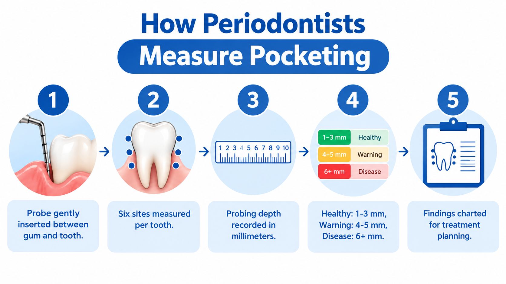
A patient often hears about pockets during a routine cleaning, then sees the hygienist write down numbers around several teeth. The term can sound ominous, but the pocket is the space between the tooth and the gum. Clinicians measure it because inflammation can loosen that seal and change the shape of the tissue around the tooth. In a healthy mouth, that space is usually 1 to 3 mm.
Why the number alone isn't the whole story
A reading tells part of the story. A 5 mm site with swollen gums may still have an inflammatory component that can improve with care, while the same reading with recession and attachment loss points to support loss around the tooth. Periodontists read that number alongside bleeding, gum position, and the full periodontal chart, and the American Dental Association describes pocket depth as one part of evaluating periodontitis.
Practical rule: treat the pocket number as a measurement that directs the exam rather than a conclusion in itself.
For adults across Wilmington, Newark, Pike Creek, Hockessin, Bear, Middletown, and Dover, that distinction matters. It keeps mild irritation from sounding like a disaster, and it keeps real disease from being dismissed as “just a little bleeding.” The next step is usually a careful periodontal exam, with probing, bleeding checks, and imaging when needed.
Pocketing in teeth is one clue inside a larger pattern. When Delaware periodontists review that pattern, they look for how bleeding, attachment loss, and bone changes fit together, then use tools such as 3D imaging to see what the eye cannot. That broader view helps them decide whether tissue is irritated, damaged, stable, or at risk of worsening.
How a Periodontal Pocket Forms
A pocket begins when gum tissue stops fitting tightly around the tooth. Plaque that lingers near the gumline lets bacterial biofilm trigger inflammation, and the body's response can start to pull the tissue attachment downward along the root. The pocket gets deeper as that attachment shifts.
The change is gradual, but the structure of the problem is serious. The fibers that help anchor the gum to the tooth, including the junctional epithelium and the periodontal ligament, begin to break down. As that support weakens, the space around the tooth becomes a pocket where bacteria and debris gather more easily, and cleaning that area becomes harder.
What changes under the gumline
Bleeding on probing often appears because inflamed tissue is fragile. Bone can also resorb beneath the gums, so pocket depth by itself does not show the whole picture. A tooth may still look fairly normal above the gumline while the support underneath is changing.
The pattern is easier to understand with the healthy gum levels and gingivitis guide in mind. Early inflammation may improve when the source is controlled. Once attachment loss and bone loss appear, the issue has moved beyond irritation and into structural change.
A deeper pocket is not just a bigger number. It often means the tooth's support system has changed.
That process develops over time. Patients are often surprised when a routine visit reveals a more advanced problem than they expected, because the pocket measurement is only the visible clue. The deeper story is in the bleeding, attachment loss, and bone changes that periodontists look for when they evaluate the tooth and decide what needs to happen next.
How Periodontists Measure and Diagnose Pocketing
A patient may hear that a tooth has “pockets” and assume the whole diagnosis rests on one number. In practice, periodontists read that number alongside bleeding, attachment loss, and bone changes, the way a clinician reads a blood pressure reading alongside symptoms and exam findings.
The measurement starts with a calibrated periodontal probe. The probe is placed from the gum margin to the base of the pocket, and the depth is recorded in millimeters at six sites per tooth, not just one: mesio-buccal, mid-buccal, disto-buccal, mesio-lingual or palatal, mid-lingual or palatal, and disto-lingual or palatal (the six recommended probing sites per tooth).
Probing depth versus attachment level
Probing depth shows the current space under the gum. Clinical attachment level shows how much support has been lost over time, because it is measured from the cementoenamel junction to the base of the pocket (the difference between probing depth and clinical attachment level). That distinction matters when the gums are swollen or when the gumline has receded. A site can look only mildly deep while hiding real support loss, and swollen tissue can make a pocket read deeper than the underlying attachment loss alone would suggest.
Diagnosis also includes bleeding, recession, tooth mobility, and imaging. The 2017 periodontitis classification staging criteria emphasize the pattern of sites at 4 mm and 6 mm or deeper, along with attachment-loss thresholds and bleeding, rather than one highest reading (the 2017 periodontitis classification staging criteria). That matters because periodontists are looking for a pattern, not a single outlier.
| Periodontitis Severity at a Glance | Interproximal Attachment Loss | Probing Depth | Other Findings |
|---|---|---|---|
| Mild | At least 3 mm at two or more sites | At least 4 mm | Bleeding may be present, distribution matters |
| Moderate | At least 4 mm at two or more sites | At least 5 mm | More support loss, often more sites involved |
| Severe | At least 6 mm plus at least one 5 mm or deeper pocket | At least 5 mm at one or more interproximal sites | Greater risk of mobility and bone loss |
A table like this shows why pocketing in teeth is only one clue in a larger story. Delaware periodontists also evaluate whether a tooth has started to move, since mobility can signal that the supporting bone and ligament need closer attention (tooth mobility assessment).
Why Pocket Depth Numbers Matter for Your Treatment Plan
A pocket depth reading is one clue, not the whole diagnosis. It helps a clinician decide what can still be managed with home care and what needs treatment below the gumline. Sites deeper than 5 mm are harder to clean thoroughly with routine brushing and flossing, and deeper pockets are more likely to stay inflamed (NCBI Bookshelf). That does not automatically mean surgery. It does mean the site needs instrumented debridement and closer follow-up than a 3 mm sulcus.
How the numbers guide decisions
Pocket depth matters because it sits alongside bleeding, recession, attachment loss, and bone change. In population data, about 42% of U.S. adults had at least one site with a probing depth of 4 mm or more, and about 88% had attachment loss of at least 3 mm at one or more sites (CDC report). Those numbers show why a few deep spots cannot be brushed aside. Deep sites are common, but common does not mean harmless.
A treatment plan usually turns on four questions:
- How deep are the pockets? Depth shows how difficult the area will be to clean and monitor.
- Is there attachment loss? Attachment loss shows whether the support around the tooth has already been damaged.
- Is bleeding still present? Bleeding points to inflammation that is still active.
- Is bone loss visible? Bone changes can shift care from cleaning alone toward more advanced treatment.
| Pocket Clues | What They May Suggest |
|---|---|
| Deep pocket with bleeding | Active inflammation that needs closer attention |
| Deep pocket with recession | Likely attachment loss, not just swollen tissue |
| Shallow pocket with bone loss | Past damage may be hidden behind a modest reading |
| Reproducible deep site in the same place | Local anatomy or plaque retention may be keeping that site unstable |
A site that keeps measuring deep in the same place is often telling a larger story. The pocket is one chapter, while bleeding, attachment loss, and bone change are the rest of the page.
i-CAT 3D cone beam imaging helps in selected cases because it shows bone contours and defects that a probe cannot reveal. Probing measures the soft tissue pocket, but imaging lets the clinician see what is happening under the surface. That matters when a site keeps bleeding or does not respond as expected.
Delaware periodontists use that combined picture to decide whether a site needs monitoring, nonsurgical therapy, surgery, or regenerative care. The reading itself is only one piece, and tooth mobility assessment can add another. A tooth that starts to move may be signaling that the supporting ligament and bone deserve closer attention.
Non-Surgical and Surgical Options for Pocketing in Teeth
A patient may hear that a pocket is “deep,” but the number alone does not decide treatment. Periodontal care starts with reducing the bacteria and inflammation that keep the pocket active. Scaling and root planing is often the first step, because it removes plaque and tartar from below the gumline and smooths contaminated root surfaces so the tissue has a better chance to settle down (NIDCR).
How treatment choices differ
Nonsurgical therapy is usually used first when the site can still be reached well enough for cleaning. Surgery comes into the picture when deep areas keep bleeding, when deposits sit in places instruments cannot fully access, or when bone and tissue changes need direct visualization. In those cases, flap surgery lets the clinician clean more thoroughly, and regenerative procedures may be considered when supporting tissue loss calls for rebuilding. guided tissue regeneration
The practice's guided tissue regeneration resource fits that same decision space. Regeneration is not used for every site, but when the defect shape and surrounding anatomy make it suitable, the goal is to support the tooth more effectively than cleaning alone can do.
Good periodontal care is sequenced. It starts with control of infection, then moves to the least invasive option that still fits the anatomy.
Laser-based treatment is covered in the next section, including when it fits and when it does not. Periodontal Associates of Delaware also offers periodontal evaluations, scaling and root planing, LANAP, traditional periodontal surgery, and regenerative procedures as part of a broader treatment menu. The right choice depends on pocket depth, bleeding, bone loss, tooth stability, and whether the site is still maintainable.
Patients often want a single answer, but periodontal therapy rarely works that way. A shallow pocket that is clean and stable may need maintenance only. A deep site with persistent bleeding may need more than home care. A tooth with structural loss may need surgery, regeneration, or both. The treatment plan follows the tissue, not the other way around.
How LANAP Laser Gum Surgery Works and When It Fits
LANAP is a laser-assisted protocol used in periodontal treatment when the anatomy and disease pattern fit the approach. The protocol uses the PerioLase MVP-7 and is designed to target diseased tissue while preserving healthy structures around the tooth. For anxious patients, that targeted approach can feel more manageable than a broad surgical experience, especially when the clinical goal is to control infection and improve pocket stability.
What patients usually need to understand
A laser does not replace the need for diagnosis. It also does not make every case a laser case. A clinician still has to decide whether the pocket pattern, bone loss, and tissue condition make the approach appropriate, or whether conventional surgery or regenerative treatment will be more useful.
A 2025 clinical-practice guideline review reported that adjunctive photodynamic therapy may offer a moderate benefit, but also noted limited evidence and a need for more long-term study. The same review found no established net benefit for several non-photodynamic laser approaches when added to scaling and root planing (2025 guideline review). A small 2025 study of 25 patients with 4 to 7 mm pockets reported improved periodontal measures after both scaling and root planing alone and combined diode-laser treatment over three months, but the short follow-up limits durable conclusions.
That's why the conversation is less about marketing and more about fit. Some pockets may respond well to meticulous nonsurgical care plus maintenance. Others may need surgery, grafting, or a combination approach. If laser treatment is considered, the key question is whether it matches the defect, the bone pattern, and the patient's long-term maintenance ability.
Important point: early pocket improvement doesn't prove long-term control.
Patients in Wilmington, Newark, and Dover often want to know whether a laser approach is “better.” The better question is whether it's appropriate for the specific tooth and the surrounding bone. That answer depends on the chart, the imaging, the bleeding pattern, and the clinician's judgment.
Maintenance and Home Care That Protect Your Results
Pocket treatment only holds if inflammation stays under control. That's why periodontal maintenance visits are usually scheduled more often than routine cleanings for people with a history of pocketing, so the team can recheck probing depths, bleeding, and attachment levels at reproducible sites. The chart matters because it shows whether a site is stable, improving, or slipping back.
What daily care has to do
Home care still does most of the quiet work. Brushing along the gumline, cleaning between teeth, and keeping plaque off the roots reduce the bacterial load that feeds inflammation. A good maintenance plan isn't just about cleaning the teeth that are easy to reach, it's about protecting the sites that got deep in the first place.
The coordinated plan often works best when the general dentist and the periodontal team keep exchanging findings. That way, changes in the chart don't get missed between visits, and persistent bleeding pockets get rechecked instead of being assumed stable. For patients in the Wilmington, Newark, and Dover offices, that coordination helps keep the follow-up consistent across locations.
Skipping maintenance is one of the fastest ways for a controlled pocket to become unstable again.
A practical sign of stability is that the same sites keep looking similar over time. If a previously deep area stops bleeding and its attachment level holds steady, that's meaningful progress even if the pocket doesn't become perfectly shallow. The goal is not cosmetic perfection, it's keeping the tooth supported and the inflammation quiet.
Patients sometimes think the problem ends when the deep cleaning or surgery ends. It doesn't. Periodontal disease is managed over time, and maintenance is the part that protects the work already done.
Common Misconceptions and Your Next Step
One common myth is that every deep pocket means a tooth is about to fall out. That's not true. A deep site can still be managed if the clinician controls inflammation, monitors attachment, and treats the supporting bone and tissue appropriately. Another myth is that bleeding gums are normal. Bleeding usually signals inflammation, and it deserves attention rather than dismissal.
What readers often misunderstand
Treatment success is not always measured by making every pocket shallow. In real periodontal care, success often means stopping progression, improving cleansability, reducing bleeding, and preserving the tooth for as long as possible. A stable 5 mm site can sometimes be more valuable than an unstable shallow one, depending on the surrounding support and how the tissue responds over time.
Patients also worry that surgery is the only path. It isn't. Some people improve with nonsurgical therapy and maintenance alone, while others need regenerative procedures, conventional surgery, or a laser-based option when the case fits. The right answer depends on the exam, not on a slogan.
What matters most is getting the pocket interpreted correctly. That means looking at the number, the bleeding pattern, the attachment level, and the bone changes together, then deciding what the tooth needs now, not what sounds most dramatic.
Individual results vary, and every mouth needs its own periodontal assessment. A board-certified periodontist can explain whether the pocket is a sign of irritation, attachment loss, or active disease that needs treatment.
If pocketing in teeth has come up in your chart, the next step is a periodontal evaluation that looks at the whole picture, not just one number. Periodontal Associates of Delaware offers evaluation, scaling and root planing, LANAP, surgery, and regenerative options across Wilmington, Newark, and Dover, so treatment can be matched to the site and the tissue involved. Request an appointment at the nearest office through Periodontal Associates of Delaware to get a clear plan for your gums and bone.