A dentist points to a dark area beside a tooth on an X-ray and mentions bone loss, a deep gum pocket, or a possible periodontal consultation. The words can land heavily. Many adults immediately worry about two things: Will the treatment hurt, and can the tooth still be saved?
Guided tissue regeneration, often called GTR, may be one option for certain periodontal defects. It isn't a magic covering or a guaranteed way to rebuild everything that gum disease destroyed. It's a carefully planned procedure that gives the body a protected space to rebuild some of the structures supporting a tooth. The right choice depends on the defect, the tooth, overall health, daily plaque control, and the patient's willingness to protect the area during healing.
Table of Contents
- When the Dentist Says You Have Lost Bone Around a Tooth
- What Guided Tissue Regeneration Actually Does
- Which Defects and Which Patients Are Typical Candidates
- Membranes, Grafts, and Biologics in Plain Language
- How the Procedure Actually Unfolds
- What the Evidence Says About Real-World Outcomes
- Risks, Recovery, and When GTR May Not Be the Right Call
- Your Next Step Toward a Treatment Plan
When the Dentist Says You Have Lost Bone Around a Tooth
Bone loss around a tooth doesn't always cause obvious symptoms. A patient may feel no pain while inflammation slowly damages the bone, periodontal ligament, and gum attachment that help hold the tooth in place. The first warning may come during a routine cleaning, when a hygienist records a deeper pocket, or when a dentist notices an irregular area on an X-ray.
That moment can feel personal and frightening. The patient may picture a long surgery, a loose tooth, or an immediate extraction. A periodontal evaluation replaces that uncertainty with details: where the pocket is, how much support remains, whether the tooth moves, and what kind of bone shape exists around the root.
A deep pocket doesn't automatically mean that GTR is needed. It means the tooth deserves a closer look at the disease, the anatomy, and the available treatment choices.
Guided tissue regeneration is one of several procedures a periodontist may consider. The technique aims to protect a cleaned defect so slower-forming periodontal cells have an opportunity to repopulate the area. Depending on the situation, treatment may instead involve non-surgical periodontal therapy, traditional periodontal surgery, laser gum treatment, soft-tissue grafting, extraction, or an implant plan.
A useful way to think about GTR is as a chance to improve support, not a promise to recreate the original tooth-supporting system. The procedure may help produce new bone, cementum, and periodontal-ligament attachment in a favorable defect. It may also leave a residual pocket, recession, or area that needs long-term maintenance.
For adults in New Castle and Kent Counties, including Wilmington, Newark, Pike Creek, Hockessin, Bear, Middletown, Dover, and nearby communities, the most helpful first step is a focused conversation with a periodontist. Clear expectations make surgical decisions less intimidating.
What Guided Tissue Regeneration Actually Does
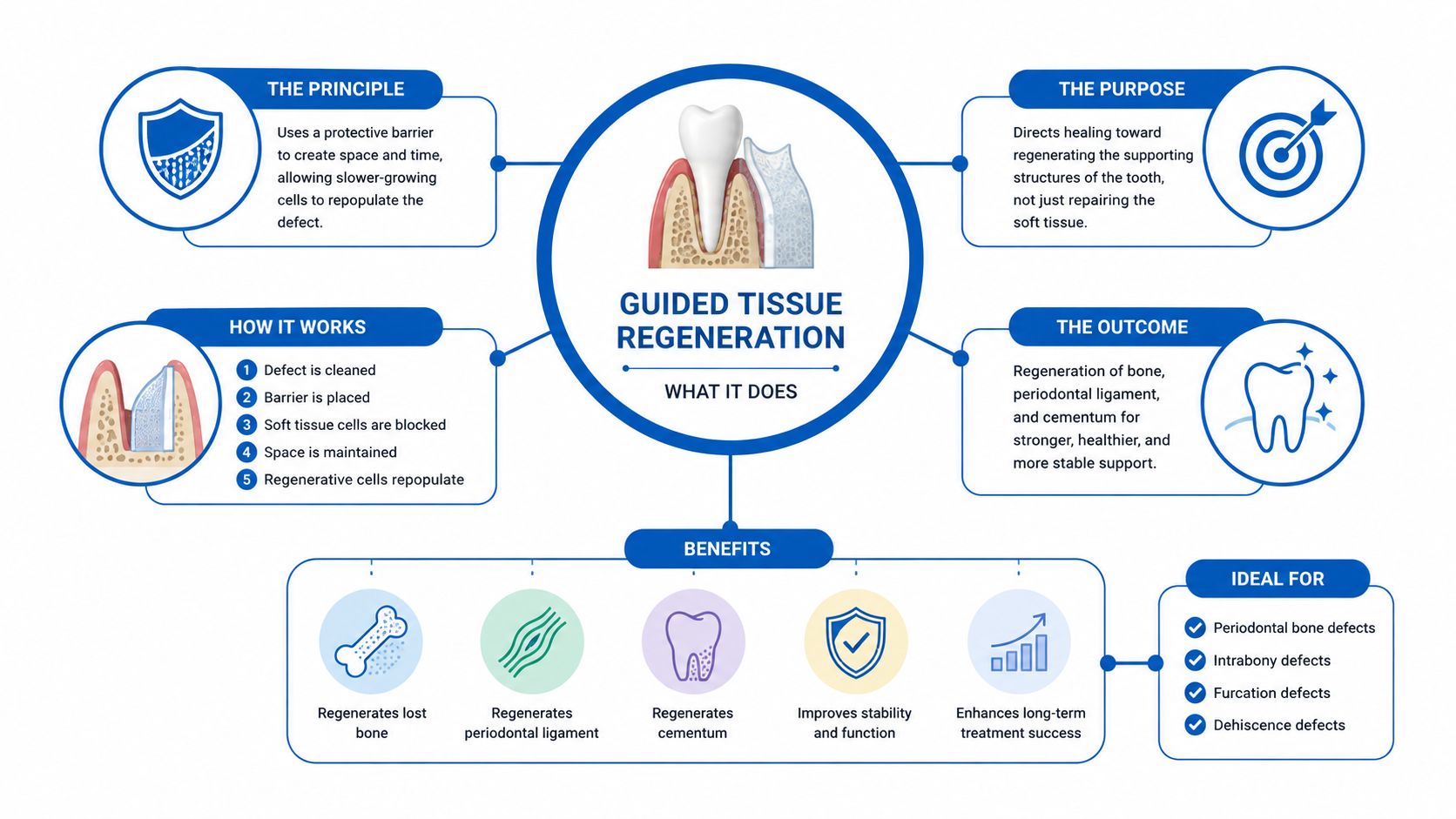
A tooth is held in place by more than the visible crown. Bone surrounds the root, a thin periodontal ligament connects the root to bone, and gum tissue forms a protective seal around the tooth. Periodontal disease can damage these supports and leave a vertical defect beside the root.
The challenge begins after diseased tissue is removed. Several cell types can enter the healing area, but they don't all build the same structures. Fast-moving gum epithelial and connective-tissue cells can fill the space before the slower periodontal-ligament and bone-forming cells arrive.
GTR uses a barrier membrane to influence that competition. A simple analogy is a small tent at a damaged campsite. The clinician cleans the site, sets a membrane over the defect, and closes the gum over it. The membrane helps maintain a protected space, much like a tent frame keeps fabric from collapsing, while the cells needed for periodontal healing gradually move into the area.
Regeneration is different from repair
Regeneration means forming new periodontal structures, including new cementum on the root, new periodontal-ligament fibers, and new bone. In the foundational human report by Sture Nyman and colleagues, published in 1982, microscopic examination after approximately three months showed new connective-tissue attachment with collagen fibers inserting into newly formed cementum. The reported attachment extended approximately 7 mm beyond a reference notch, while bone regeneration reached the existing bony crest. The report is described as an early milestone in modern periodontal regeneration in this historical review of guided tissue regeneration.
Repair can produce a different result. Fast-growing tissue may seal the root with a long junctional epithelium, making the area healthier or easier to maintain without recreating the original attachment apparatus. That improvement can still matter clinically, but it isn't the same as true histologic regeneration.
Before recommending treatment, Periodontal Associates of Delaware may use i-CAT 3D cone beam imaging along with probing and other records to understand the defect's shape. Three-dimensional information can help show whether the remaining bone walls can contain the healing space. It can't predict the result by itself, but it adds important information to the clinical examination.
Which Defects and Which Patients Are Typical Candidates
A tooth may have lost bone in a narrow vertical pocket, while the surrounding bone remains partly intact. That shape gives GTR a contained space to protect during healing. A deeper defect with several remaining bony walls is generally more favorable than a broad, shallow defect with little bone around it. The procedure is therefore defect-dependent, not a guaranteed response to bone loss.
Periodontal literature identifies intrabony defects deeper than 3 mm and selected Class II furcation defects as situations in which membranes may provide clinically relevant, relatively predictable regenerative results, although complete resolution is uncommon. A furcation defect lies between the roots of a multi-rooted tooth. Remaining bone in that area can help stabilize the clot and membrane while the wound heals. These indications are summarized in the periodontal regeneration review.
The anatomy matters, but it isn't the only factor
A periodontist assesses how well the defect suits the procedure rather than relying on one X-ray. The evaluation may include:
- Defect walls: Remaining walls can help contain a membrane, graft, or blood clot.
- Inflammation: Active infection and uncontrolled bleeding can disturb wound stability.
- Tooth stability: Mobility, root shape, biting forces, and remaining support affect the treatment plan.
- Furcation involvement: Selected furcation defects may respond differently from defects around a single-rooted tooth.
- Plaque control: The patient must be able to keep the mouth clean and protect the surgical site.
- Medical history: Smoking, diabetes control, medications, and healing capacity can alter risk.
- Maintenance commitment: Any regenerated or repaired support still requires professional periodontal care.
A good candidate has a defect that can be contained and a healing environment the patient can protect. Excellent imaging cannot compensate for plaque control the patient cannot maintain. A similar defect may heal less predictably when the surgical site is repeatedly disturbed, inflammation remains active, or aftercare is difficult to follow.
The decision also involves trade-offs. A contained defect may offer a reasonable opportunity for regeneration, but the procedure still requires surgery, healing time, careful cleaning, and ongoing maintenance. Some defects are too shallow, too open, difficult to close, or unlikely to stay clean. In those situations, periodontal Associates of Delaware should discuss whether regenerative treatment offers enough likely benefit to justify its burden, rather than presenting GTR as the automatic choice.
Membranes, Grafts, and Biologics in Plain Language
Patients may hear several material names during a consultation. These products don't all do the same job, and adding more materials doesn't automatically create better regeneration.
A resorbable collagen membrane gradually breaks down in the body, so it usually avoids a second procedure for removal. A nonresorbable e-PTFE membrane can maintain space effectively, but it generally requires later removal and creates a greater management burden if it becomes exposed. The choice depends on the defect, closure, access, and the clinician's treatment plan.
A graft may be placed beneath a membrane when the defect needs additional structural support. Graft categories include material from the patient's own body, donated human tissue, animal-derived material, or synthetic substitutes. A biologic, such as an enamel-matrix derivative, platelet concentrate, or growth-factor product, may be selected to support a particular healing goal. None should be treated as a universal “best” option.
What each material contributes
| Material | What It Does | Typical Reason It Is Chosen |
|---|---|---|
| Resorbable collagen membrane | Excludes rapidly migrating cells and supports wound stability as it breaks down | Avoids planned membrane-removal surgery |
| Nonresorbable e-PTFE membrane | Maintains a protected regenerative space | Provides firm space maintenance when closure and follow-up can be managed |
| Bone-replacement graft | Adds scaffold or structural support inside a defect | Helps stabilize space in less-contained anatomy |
| Enamel-matrix derivative | Supports a biologic regenerative environment | May be considered when the defect and healing conditions are appropriate |
| Platelet concentrate or growth-factor product | Provides biologic components intended to support healing | Selected selectively according to anatomy and treatment goals |
A randomized study of infrabony defects with unfavorable architecture found that a collagen membrane combined with decalcified freeze-dried bone allograft produced a mean probing-pocket-depth reduction of 4.06 mm and clinical attachment-level gain of 3.54 mm at six months, compared with 3.20 mm and 2.50 mm for membrane treatment alone, respectively. The findings support the idea that a graft may add space support in selected defects, but they don't mean every patient needs one. The study is reported in this clinical trial of membrane and graft treatment.
Patients who want more background on graft materials can review the practice's periodontal bone graft resources. The final selection should still follow an examination, not a product list.
How the Procedure Actually Unfolds
The unfamiliar parts of surgery often cause more anxiety than the procedure itself. A clear sequence helps patients understand what happens before, during, and after GTR.
Before surgery
The periodontist records pocket depths, bleeding, attachment levels, tooth mobility, furcation involvement, and the condition of the surrounding gums. Imaging may include i-CAT 3D cone beam imaging when the defect's three-dimensional shape needs closer evaluation. Treatment planning may also involve communication with the general dentist or another specialist so the tooth's restorative and bite-related needs are included.
The consultation should answer practical questions. What structure remains? What would success look like? Which material is being considered? What happens if the membrane becomes exposed? How will the tooth be maintained after healing?
On the treatment day
Local anesthesia numbs the area. The periodontist makes a carefully planned flap to see the root and defect, removes diseased deposits and tissue, and prepares the root surface. A membrane is positioned over the defect, with graft material added when the anatomy calls for extra support. The gum is then repositioned and sutured for primary, tension-free closure.
Many patients feel pressure, movement, or vibration rather than sharp pain during the procedure. The practice may emphasize minimally invasive approaches when appropriate, with the aim of limiting postoperative discomfort and recovery demands. The exact surgical approach depends on access, defect shape, and the structures that need treatment.
After the appointment
The early days require protection, not forceful cleaning. Patients may receive instructions for a modified diet, careful brushing away from the surgical site, short-term antimicrobial rinses, and other prescribed care. Suture removal and follow-up visits allow the periodontist to check closure and healing.
A regenerative result also depends on the maintenance phase. Ongoing periodontal maintenance, often scheduled every three to four months, helps control inflammation and plaque around the treated tooth and throughout the mouth.
What the Evidence Says About Real-World Outcomes
The core patient question is not just whether GTR “works.” It's whether the procedure offers enough likely benefit for a particular defect to justify surgery, healing restrictions, exposure risk, and long-term maintenance.
A recent systematic review and meta-analysis examined 17 randomized controlled trials involving 501 periodontal intrabony defects. Follow-up ranged from 5 to 20 years, with a mean follow-up of 9.32 ± 4.75 years. Across studies with at least five years of follow-up, GTR-treated defects showed an average clinical attachment-level gain of 3.27 mm, with a 95% confidence interval of 2.90 to 3.65 mm, and probing-depth reduction of 4.04 mm, with a 95% confidence interval of 3.69 to 4.38 mm, compared with baseline. These figures come from the long-term systematic review and meta-analysis of GTR outcomes.
In patient language, a smaller probing depth may make an area easier to clean and monitor. Clinical attachment gain reflects improved measured support around the tooth. Those changes can be meaningful, but an average doesn't predict exactly what will happen around one person's tooth.

Improvement from baseline is not superiority
The review rated the certainty of evidence as low for clinical attachment and probing-depth outcomes because of study bias and inconsistencies. It also did not conclusively establish that GTR is superior to conventional open-flap debridement over the long term.
Earlier evidence involving furcation defects found greater bone fill, attachment gain, and furcation closure with GTR than with open-flap debridement, particularly in mandibular molars. Combining GTR with an osseous graft generally produced stronger outcomes than GTR alone in that earlier evidence, but treatment decisions still need to match the defect.
A radiograph can show bone fill, but an image alone doesn't prove that the original periodontal attachment apparatus has been recreated. Histologic regeneration requires new cementum, periodontal-ligament fibers, and bone in the proper relationship. Patients comparing procedures can also review the practice's information about gum graft failure rate, while remembering that gum grafting and GTR address different clinical problems.
Risks, Recovery, and When GTR May Not Be the Right Call
An honest consent conversation includes the possibility that the membrane may become visible through the gum or that the flap may separate. Exposure can allow bacterial contamination, compromise the protected space, and shift healing toward repair rather than the intended regenerative outcome.
A systematic review of early healing found membrane exposure or flap dehiscence in approximately 26% of GTR-treated two- to three-wall intrabony defects, compared with 6.5% in enamel-matrix-derivative treatment. The comparison is reported in this systematic review of early periodontal regenerative healing. These figures don't predict an individual result, but they explain why closure, hygiene, and follow-up deserve serious attention.
Other possible concerns include swelling, sensitivity, temporary or lasting recession, infection, persistent pocketing, and less bone or attachment gain than hoped. A periodontist may reduce risk through careful selection, meticulous debridement, stable closure, smoking-cessation support, and close postoperative monitoring.
When another treatment may make more sense
GTR may not be the right call when active inflammation has not been controlled, the defect cannot contain a membrane, the tooth lacks enough remaining support, or the patient cannot protect the site during healing. A different approach may better match the problem:
- Active periodontal infection: LANAP laser gum surgery using the FDA-cleared PerioLase MVP-7 may be considered as part of a plan to control disease.
- A tooth that cannot be predictably saved: Extraction followed by implant planning may be discussed, with X-Nav dynamic surgical navigation used for selected implant procedures.
- Gum recession or exposed roots: Soft-tissue grafting addresses coverage and tissue thickness rather than rebuilding a vertical bone defect.
- A maintainable defect with less surgical risk: Non-surgical periodontal therapy or conventional access surgery may be more appropriate.
Smoking, diabetes control, plaque levels, tooth mobility, defect anatomy, and commitment to maintenance can all change the balance. The best plan isn't the one with the most materials. It's the one whose potential benefit justifies its risks for the tooth and the patient.
Your Next Step Toward a Treatment Plan
Guided tissue regeneration may provide meaningful improvement when a defect has favorable anatomy and the patient can support healing. It isn't a universal solution, and no responsible clinician can promise complete reconstruction, a guaranteed cure, or preservation of every tooth.
Periodontal Associates of Delaware is a board-certified periodontal practice with diplomate status from the American Board of Periodontology. Care is delivered across three Delaware offices in Wilmington, Newark, and Dover, with collaboration among general dentists and specialists serving New Castle and Kent Counties.
A consultation may include periodontal charting, medical-history review, tooth-stability assessment, and imaging such as i-CAT 3D cone beam imaging. For implant treatment when a tooth cannot be saved, the practice also uses X-Nav dynamic surgical navigation. Individual results vary, so each patient should consult a periodontist for advice suited to the mouth, health history, defect anatomy, and treatment goals.
Periodontal Associates of Delaware evaluates periodontal bone loss and can determine whether guided tissue regeneration, another regenerative approach, disease-control treatment, or implant planning fits the tooth's condition. Adults in Wilmington, Newark, Dover, and surrounding Delaware communities can request an appointment with Periodontal Associates of Delaware at the nearest office.