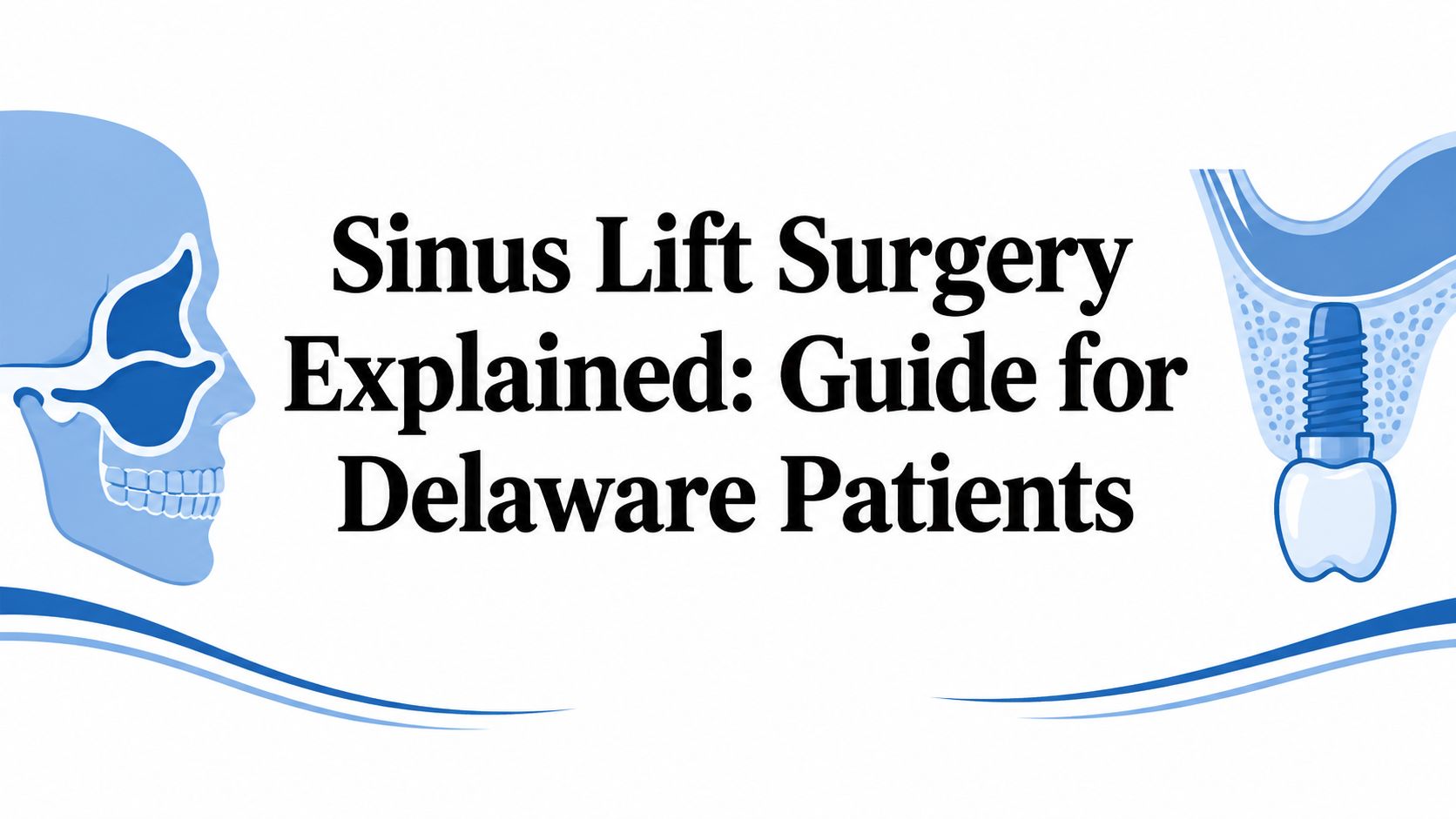
A sinus lift surgery adds bone to the upper back jaw when natural bone is too thin to support dental implants, and Periodontal Associates of Delaware offers both lateral and crestal approaches using advanced 3D imaging. The right approach depends largely on residual bone height, sinus health, implant goals, and how much healing time the patient can allow.
A person in Wilmington, Newark, or Dover may lose a back tooth, wait while life gets busy, and later learn that an implant can't be placed safely without rebuilding the area first. That news can feel discouraging, especially for adults already dealing with gum disease, recession, bone loss, or anxiety about oral surgery. A sinus lift isn't a sign that implant treatment has failed. It may be the reconstructive step that makes stable implant placement possible.
Table of Contents
- Why Many Delaware Patients Need Sinus Augmentation
- Understanding Sinus Anatomy and Bone Loss
- Lateral vs Crestal Sinus Lift Approaches
- Graft Materials, Risks, and Safety Data
- How Advanced Imaging Improves Outcomes
- Recovery Timeline and What to Expect
- Is Sinus Lift Surgery Right for You
Why Many Delaware Patients Need Sinus Augmentation
An adult in New Castle or Kent County may have a missing upper molar and expect a straightforward implant. During planning, the periodontist may find that the jawbone has narrowed or shortened, leaving the sinus too close to the proposed implant site. Instead of placing an implant into weak support, the clinician can recommend sinus augmentation, also called a sinus lift, to create additional bone volume.
Bone loss after tooth removal is common because the jaw no longer receives the same stimulation from chewing. The natural maxillary sinus, an air-filled space above the upper back teeth, can also extend downward as the supporting ridge changes. The result resembles a house whose foundation has been weakened by erosion. The house may still look intact from the outside, but it can't safely carry a new upper level until the foundation is reinforced.
Sinus lift surgery is therefore reconstructive rather than cosmetic. It lifts the sinus lining and places graft material beneath it, creating a stronger site for a future implant. The procedure became established in implant dentistry after Tatum introduced it at an Alabama dental implant conference in 1976, and it later became a standard way to increase bone height in the posterior maxilla. A review of a radiographic screening study involving 2,837 patients found that 57.43% of subantral ridge measurements were 4 mm or less, demonstrating how often significant deficiency can affect implant candidates. The clinical review of sinus augmentation history and anatomy/gendent_ja15_almas.pdf?sfvrsn) provides further context.
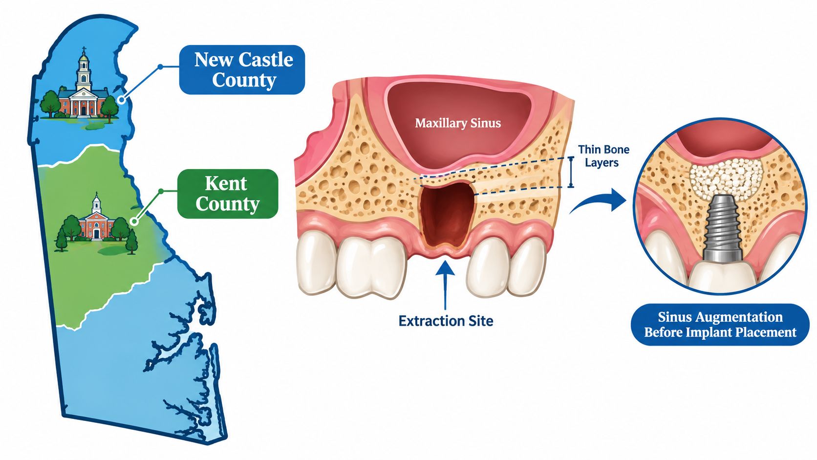
The anatomy can be more extensive than a single missing tooth suggests. In cases with 6 mm or less of residual ridge height, the same study found that atrophy extended to the second premolar region in 39.32% of cases, to the first premolar in 20.51%, and to the canine fossa in 10.84%. These findings help explain why a sinus lift is often a foundational part of posterior upper-jaw rehabilitation, not an unusual add-on.
Understanding Sinus Anatomy and Bone Loss
The maxillary sinuses sit inside the cheek area, directly above the roots of the upper premolars and molars. A thin tissue lining, called the Schneiderian membrane, separates this air space from the jawbone. When a tooth is present, its roots and surrounding bone occupy the area below the sinus. After tooth loss, that support can gradually diminish.
The process is similar to removing a tree from firm soil. The soil settles into the empty space, and the ground level drops. In the upper jaw, the ridge may lose both width and height, while the sinus floor may sit closer to the gum. An implant still needs enough surrounding bone to remain stable, so the clinician must measure the available foundation before selecting a surgical plan.
Residual bone height, often shortened to RBH, is the vertical distance between the crest of the jaw and the floor of the sinus. It isn't the only planning factor, but it gives the periodontist a practical starting point. A 3D image can show whether the ridge resembles a strong frame for a deck or a weakened frame that needs reinforcement before it can carry weight.
Practical rule: An implant shouldn't be planned from a surface view alone. The periodontist must evaluate the ridge, sinus membrane, neighboring teeth, and the proposed implant position together.
Several conditions can contribute to an inadequate site:
- Tooth loss: The jaw may shrink after extraction because it no longer receives normal functional stimulation.
- Periodontal disease: Long-standing gum infection can destroy the bone that once supported the tooth.
- Natural sinus anatomy: Some patients begin with a sinus that extends farther downward than expected.
- Delayed treatment: More time without a tooth may allow the ridge to become less suitable for direct implant placement.
A sinus lift addresses the vertical limitation beneath the sinus. Other procedures, such as ridge preservation or bone augmentation, may be appropriate when the main problem involves width or bone loss in another part of the jaw. Careful diagnosis prevents a one-size-fits-all recommendation.
Lateral vs Crestal Sinus Lift Approaches
The choice between a lateral and crestal approach usually begins with residual bone height, then expands to include sinus anatomy, the number of implants, grafting needs, and whether an implant can be stabilized at the same visit.
A lateral window approach reaches the sinus through a small opening in the side wall of the upper jaw. The clinician gently lifts the membrane and places graft material in the space below it. This method gives the surgeon broader access and is commonly favored when the posterior maxilla is severely atrophic or when a larger vertical increase is needed.
A crestal approach starts at the top of the ridge, often through the planned implant site. Gentle osteotome, hydraulic, balloon-assisted, or related techniques lift the membrane from below. Because the access point is smaller, the approach may involve less surgical disruption, but it offers less working room and can be less suitable when the ridge has very little native bone.
Consensus guidance often places the crestal approach above about 5–6 mm of available bone, while a lateral lift with simultaneous implant placement may be considered when approximately 3–4 mm remains. Consensus guidance on sinus augmentation and implant placement describes this practical relationship. Another current review notes that more than 5 mm may support a crestal osteotome approach, while bone height under 4 mm often favors a lateral window. The review of technique selection by residual bone height explains why the measurement matters.
| Feature | Lateral Window Approach | Crestal (Osteotome) Approach |
|---|---|---|
| Access point | Through a window in the side wall of the upper jaw | Through the ridge and planned implant site |
| Typical use | Severe atrophy or a larger vertical graft | More favorable residual bone height |
| Working access | Broad visualization and access to the sinus floor | More limited access through the crest |
| Surgical disruption | May involve more swelling and recovery | Often less invasive when anatomy is suitable |
| Graft planning | Commonly used when graft volume is important | May use a smaller graft volume or selected graftless methods |
| Implant timing | May be staged or combined, depending on stability | May be combined when enough native bone provides initial stability |
When RBH is around 4–6 mm, selected patients may be candidates for a one-stage lateral lift with grafting and immediate implant placement. As RBH falls below 4 mm, initial implant stability becomes more difficult, and grafting is generally more dependable than relying on a blood clot alone. Evidence on residual bone height, grafting, and one-stage treatment supports this planning principle.
The measurement doesn't dictate treatment by itself. A periodontist must also inspect membrane thickness, sinus health, ridge width, bite forces, and the implant's planned position.
Graft Materials, Risks, and Safety Data
Bone grafting material fills the space beneath the raised sinus membrane and provides a scaffold for new bone formation. The material may come from a human donor source, an animal-derived source, the patient, or a synthetic substitute. The choice depends on anatomy, treatment goals, medical history, and the clinician's assessment of how much support the future implant will need.
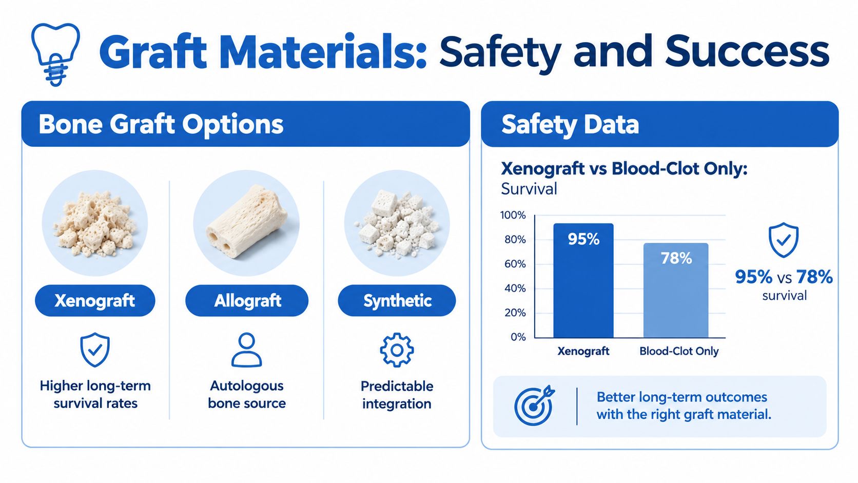
Xenografts are animal-derived materials used as a scaffold. Allografts come from a human donor source. Synthetic grafts use manufactured substitutes designed to support integration. No single material is right for every patient, and graft selection shouldn't be separated from the surgical approach or the amount of native bone remaining.
Comparative evidence has found higher implant survival with xenograft-assisted augmentation than with blood-clot-only procedures across the reported follow-up points. That distinction matters most when the patient has very limited native bone, because the implant depends more heavily on the augmented site for early stability.
What the safety data means
The Schneiderian membrane is delicate, making perforation the main intraoperative concern. Published estimates vary widely by technique and study design. Reported rates range from 0% to 58.3%, with a mean occurrence of 19.5%, while another meta-analysis reported a weighted incidence of 23.5%. Technique-specific summaries place average perforation frequencies at 15.7% to 23.1% for lateral approaches and 3.1% to 6.4% for transcrestal approaches. The systematic review of sinus lift complications and outcomes explains why these figures shouldn't be treated as a single universal risk.
Acute maxillary sinusitis has been reported in 10% to 20% of patients after sinus lift procedures, while symptoms consistent with chronic rhinosinusitis have been reported in 4% to 8%. A separate meta-analysis found 3,749 surviving implants out of 3,975, representing a 94.3% survival rate after sinus augmentation. The meta-analysis of implant survival after sinus augmentation puts those risks in the context of generally favorable implant outcomes.
A safe decision isn't based on reassurance alone. It comes from matching the patient's sinus condition, bone height, graft plan, and surgical access before treatment begins.
Patients with existing sinus disease, anatomical obstruction, or a membrane perforation may need more detailed evaluation. The relevant question isn't only whether sinus lift surgery is generally safe. It is whether the patient's individual sinus and bone conditions call for a specific workup or a different implant plan. The review of sinus conditions and patient-specific risk factors addresses that distinction.
How Advanced Imaging Improves Outcomes
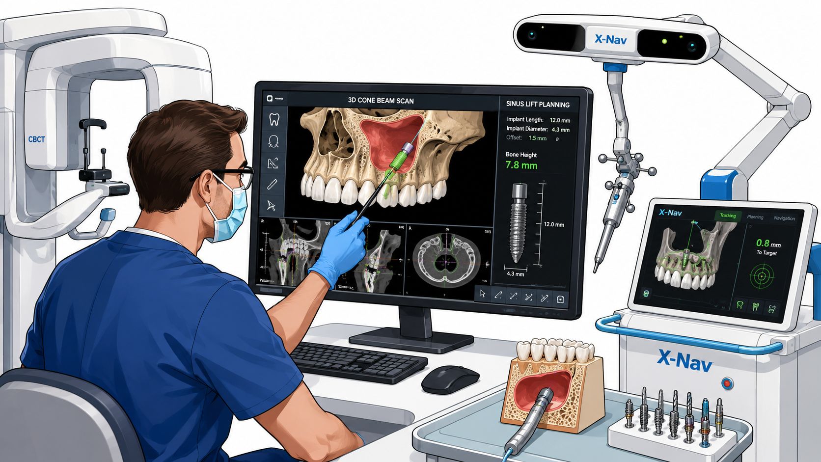
A two-dimensional dental image can suggest bone loss, but it can't show the complete three-dimensional relationship between the ridge, sinus floor, membrane, roots, and planned implant. i-CAT 3D cone beam imaging gives the periodontist a more detailed map for measuring residual bone height and identifying anatomical features that could affect the surgical approach.
At the Wilmington, Newark, and Dover offices, the practice can use i-CAT imaging as part of implant and sinus augmentation planning. The scan helps determine whether a crestal approach may be suitable, whether a lateral window offers better access, and whether the sinus appears to require additional evaluation before surgery. It also allows the planned implant position to be assessed in relation to the final restoration rather than chosen only by the existing bone.
Planning before the procedure
The clinician can review:
- Bone height and width: The scan shows the available foundation beneath the sinus and around neighboring teeth.
- Sinus anatomy: The clinician can assess the floor, internal contours, and features that may affect membrane elevation.
- Implant trajectory: The proposed path can be evaluated for stability and restorative function.
- Surgical access: The image supports a decision between a crestal, lateral, staged, or combined plan.
X-Nav dynamic surgical navigation can help guide implant placement along the planned path while the procedure is underway. Navigation doesn't remove biological risk or guarantee a particular result, but it can connect the digital plan with the surgical field and support controlled placement.
The practice also provides LANAP laser gum surgery using the FDA-cleared PerioLase MVP-7 when periodontal disease requires treatment before implant reconstruction. Controlling active gum infection may be an important part of preparing the mouth for future implant care, although LANAP isn't a replacement for sinus augmentation when the upper jaw lacks vertical bone.
Patients can review the practice's imaging and navigation systems on the technology and treatment planning page. Individual anatomy still determines whether these tools are appropriate and how they fit into a complete plan.
Recovery Timeline and What to Expect
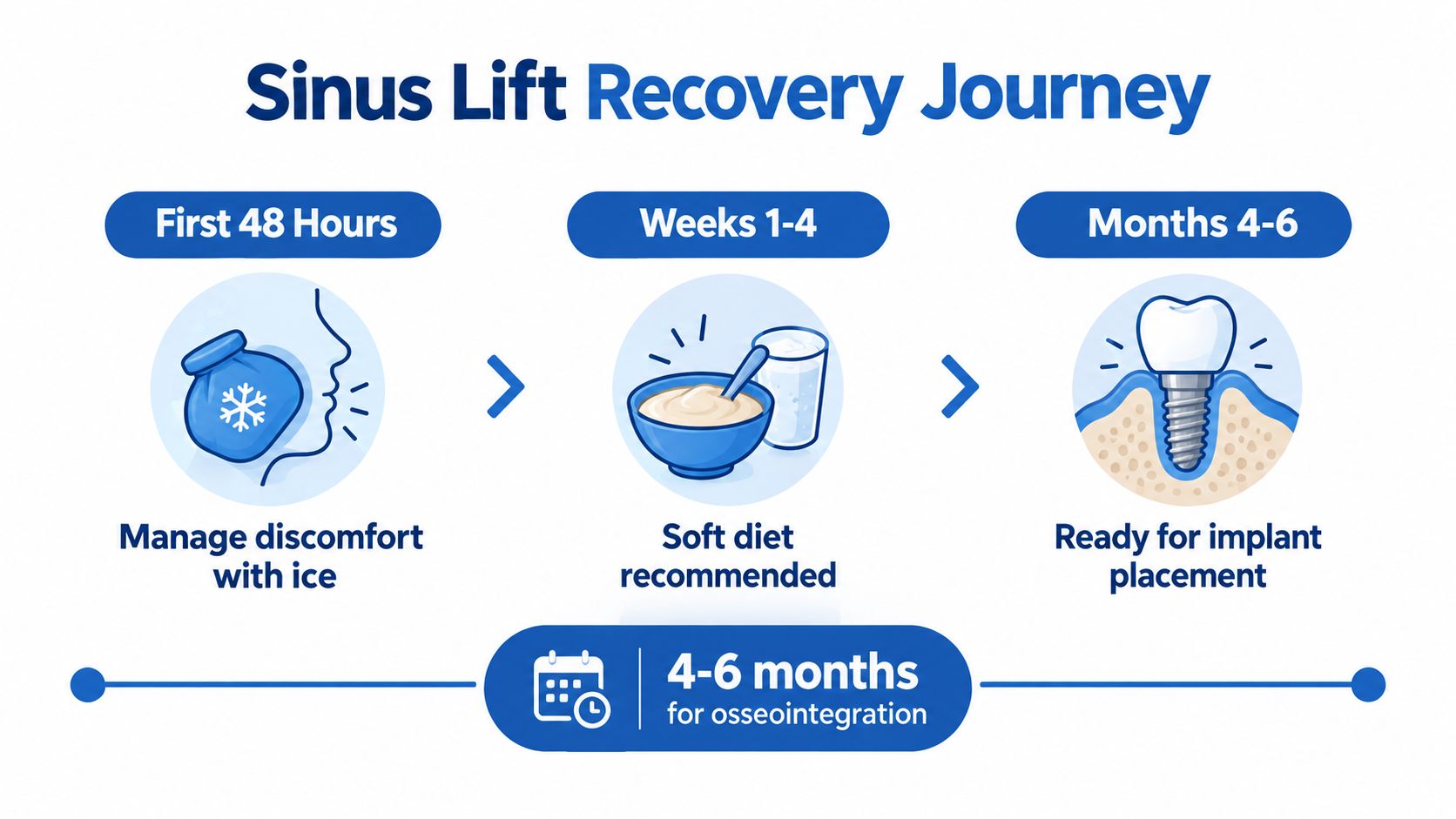
Recovery has two parts. The first involves healing of the gum tissue and surgical site. The second involves gradual bone formation and integration, which takes much longer and determines when the area can support an implant.
During the early period, swelling, bruising, pressure, and tenderness may occur. Many patients return to ordinary activities within a few days, but the appropriate pace depends on the extent of surgery, the approach used, overall health, and the clinician's instructions. A crestal procedure may involve less postoperative disruption, while a lateral window may require more time before strenuous activity feels comfortable.
Protecting the surgical site
Patients commonly receive individualized instructions such as:
- Avoid pressure changes: Don't blow the nose forcefully, and follow the clinician's guidance for sneezing.
- Use prescribed medication: Take medications exactly as directed rather than adjusting them without clinical advice.
- Choose gentle foods: A soft diet can reduce chewing pressure while the site begins to heal.
- Keep follow-up visits: The periodontist checks the tissue, graft area, and healing response.
- Limit strenuous activity: Heavy exertion may increase pressure or bleeding at the surgical site.
The graft needs time to become part of the supporting bone. A review of osteotome-mediated sinus elevation reported a mean osseointegration period of 4.9 months, with a maximum height gain of 4.62 mm, a minimum gain of 2.07 mm, and a reported success rate of 95.5%. The review of bone gain and integration after sinus elevation provides the clinical context for this waiting period.
When to call the periodontist
Increasing rather than improving pain, persistent bleeding, worsening swelling, fever, foul drainage, or concerning nasal symptoms should prompt a call to the dental team. A patient shouldn't wait for a scheduled visit when symptoms appear unusual or intensify.
The sinus augmentation recovery resource can help patients prepare questions and understand routine care instructions. It doesn't replace personalized postoperative guidance, because healing varies from one surgical site to another.
Is Sinus Lift Surgery Right for You
Sinus lift surgery may be appropriate when the upper back jaw lacks enough vertical bone for predictable implant support. The decision becomes less straightforward when the patient has active periodontal infection, sinus symptoms, poor healing factors, insufficient ridge width, or a treatment timeline that can't accommodate graft integration.
A board-certified periodontist should evaluate three broad questions:
- Is there enough healthy tissue to begin? Active gum disease and untreated infection may need attention before augmentation or implant placement.
- What does the 3D anatomy permit? Residual bone height, sinus shape, membrane condition, and ridge width help determine whether a lateral or crestal approach is more suitable.
- Can the patient protect the graft during healing? Following medication, diet, activity, and follow-up instructions is part of the treatment, not an optional extra.
The procedure has a strong clinical track record, but outcomes aren't identical for every starting condition. Evidence has found a major difference between studies involving residual bone height of at least 4 mm, where cumulative survival was reported as 100%, and studies involving less than 4 mm, where survival was 69.6%. Those findings don't predict an individual result, but they show why a bone-height measurement should guide the plan rather than serve as a minor detail.
A periodontist may also recommend treating gum disease before implant reconstruction. The practice's LANAP protocol with the FDA-cleared PerioLase MVP-7, along with i-CAT imaging and X-Nav navigation when appropriate, can support a coordinated periodontal and implant plan across Wilmington, Newark, and Dover. The sinus augmentation cost and planning resource can help patients prepare financial questions without assuming that every case has the same treatment sequence or coverage.
Individual results vary, and only a periodontist can determine suitability after an examination, medical review, and appropriate 3D imaging. A consultation should leave the patient with a clear explanation of the available bone, the recommended approach, graft material, risks, recovery expectations, and alternatives.
Patients with missing upper back teeth and limited bone can request a sinus augmentation evaluation, 3D imaging review, and implant planning with Periodontal Associates of Delaware. The board-certified periodontal practice serves Wilmington, Newark, Dover, and surrounding Delaware communities, and appointments can be requested at the nearest office through the appointment request page.