An implant may be placed on the same day as extraction when the socket and bone allow it, or after a healing period ranging from about 4 weeks to more than 6 months when they don't. But if the gum looks healed, does that mean the bone underneath is ready?
That distinction explains why implant timing isn't just a countdown after tooth removal. Gum closure, bone repair, infection control, and the ability to hold an implant firmly are different parts of healing.
For adults wondering how long after extraction can you get an implant, the most useful answer includes both a possible date and the clinical conditions needed to reach it. Waiting can be active treatment, especially when a periodontist is managing infection, preserving bone, or preparing thin gum tissue.
Table of Contents
- Implant Timing Options at a Glance
- Same-Day Implants and When They Fit
- Waiting Periods After Tooth Removal
- Clinical Factors That Change Your Timeline
- Imaging, Grafting and the Question of Resetting the Clock
- Gum Disease and Recession Before Implant Surgery
- Book a Timing Consultation at Our Nearest Office
Implant Timing Options at a Glance
What separates a same-day implant from one placed months later? The answer is what the site can support at each stage of healing. The socket works like a foundation. Its walls, bone volume and freedom from infection determine when that foundation can support an implant.
The ITI consensus on implant placement and loading protocols describes immediate placement, early placement after 4 to 8 weeks or 12 to 16 weeks, and late placement after complete healing, typically more than 6 months. These intervals give the periodontist a vocabulary for timing. The calendar follows biology, and that biology can be treated.
Implant timing options after extraction
| Timing option | Typical interval after extraction | What it depends on | Reported survival range |
|---|---|---|---|
| Immediate placement (Type 1) | Same day as extraction | Enough bone, intact socket walls, and primary stability | 93.8% to 100% |
| Early placement (Type 2) | 4 to 8 weeks | Soft tissue closure with some socket anatomy kept | 95% to 100% |
| Early placement (Type 3) | 12 to 16 weeks | Partial bone healing and fill | 95% to 100% |
| Delayed placement (Type 4) | More than 6 months, or longer with grafting | Complete bone healing | 92% to 100%, for studies defining delayed placement as after more than 4 months |
The survival ranges in the table come from a literature review of immediate, early, and delayed implant placement, which reported 93.8% to 100% for immediate placement, 95% to 100% for early placement, and 92% to 100% for delayed placement after more than 4 months. Its delayed category uses a shorter interval than the complete-healing definition in the consensus classification. Read ranges as summaries of study groups. They are not personal predictions, and a surviving implant can still have gum recession, implant exposure or additional treatment.
Choosing between the options
The review associated immediate placement with more complications, including gum recession and implant exposure. Early placement had fewer complications in the studies reviewed. For an individual patient, the choice still depends on the site and the planned restoration. Survival figures describe whether an implant remained in place. They leave out how the gum and bone look afterward.
Practical rule: The right appointment date is the earliest date when the site can support the planned treatment predictably.
A periodontist selects timing through examination and three-dimensional imaging. Extractions involving infection, gum disease or significant bone loss more often require a staged approach. The site may need treatment, bone grafting or soft tissue support before it can provide a suitable foundation. In that situation, the wait is part of the treatment plan, and the implant date follows the tissues rather than a fixed count of days.
Same-Day Implants and When They Fit
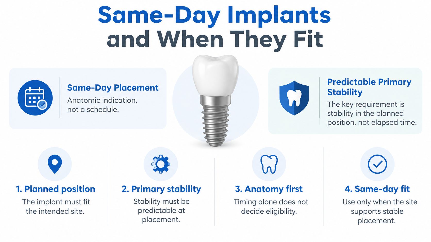
Same-day placement is an anatomical decision, not a scheduling preference. An open socket creates space, but space alone doesn't hold an implant securely. The socket works like a hole in a wall: the opening gives you room for a fixture, but the surrounding wall must grip it firmly. In clinical terms, the implant needs primary stability, meaning it is mechanically secure in bone at placement. It also needs to sit where the future crown requires, not merely where the remaining bone offers support.
Clinical requirements for immediate placement
The ITI consensus on placement in extraction sockets identifies suitable anatomy and predictable stability as central requirements. It also notes that a socket gap of 2 mm or less may heal spontaneously, while wider gaps or missing walls generally need regenerative support.
A same-day assessment considers these conditions:
- Enough remaining bone. Bone must hold the implant firmly in the intended position, including suitable bone beyond or around the socket.
- A thick, intact facial wall. This is the bone on the cheek or lip side of the socket, which helps support the visible gum contour.
- Favorable soft tissue. The surrounding gums must support the surgical and restorative plan.
- Infection status. Active infection affects the sequence, and the factors that can reset the timeline explain how it shifts placement to a later visit.
- Correct positioning for the crown. An implant that is secure but poorly positioned can create restorative and appearance problems.
A narrow gap around the implant and a missing socket wall call for different responses. Wider spaces or damaged walls may require a membrane, grafting material, or both, even when placement remains possible.
Placement and a finished crown are different milestones
An immediately placed implant still needs osseointegration, the biological process through which bone bonds to its surface. Same-day placement therefore doesn't guarantee routine use of a permanent crown that day. Extraction-related remodeling can also change the gum contour even when an implant survives, which matters especially where the gumline is visible during smiling.
At Periodontal Associates of Delaware, implant planning and placement use 3D cone beam imaging and X-Nav dynamic surgical navigation, with sub-millimeter precision. These tools support accurate planning and placement. They do not replace the need for suitable bone and healthy tissues.
For upper back teeth, the separate question of whether a sinus lift and implant can be performed together may also affect the surgical plan.
A useful consultation checklist includes:
- Stability: Can the implant be held securely in the correct position?
- Socket condition: Are the facial wall and surrounding tissues suitable?
- Backup plan: What happens if extraction reveals more damage than expected?
- Restoration: Will a temporary tooth be appropriate during healing?
Waiting Periods After Tooth Removal
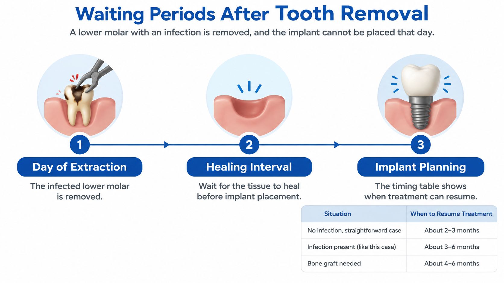
Consider an adult whose lower molar needs removal because of infection. The periodontist determines that placing an implant during the extraction appointment would be unsuitable, so treatment begins with removal of the tooth and management of the infected site.
That patient hasn't stopped implant treatment. The patient has started a staged plan designed to prepare the foundation before placement.
A healing timeline with decision points
The intervals in the timing table function as reassessment windows, not automatic surgery dates. In this lower-molar example, the sequence could look like this:
- At extraction: The clinician removes the tooth, addresses infection, examines the socket, and determines whether ridge preservation is appropriate.
- At the soft tissue checkpoint: The clinician checks closure, inflammation, and whether the available bone could now support stable early placement.
- At the partial bone-healing checkpoint: Examination and imaging assess developing bone fill and whether the implant can be positioned securely.
- After complete healing: The clinician evaluates the healed ridge and any augmentation before proceeding with a later placement plan.
Not every patient needs every checkpoint or a new scan at every visit. The periodontist chooses follow-up examinations and imaging according to the defect, treatment performed, and healing progress.
If the site still lacks support at a reassessment, the date may change. That adjustment should come with an explanation of what remains unresolved, such as persistent inflammation, insufficient bone, or a graft that needs further evaluation.
What happens between appointments
Active waiting has a purpose. The clinician may be monitoring tissue closure, checking disease control, or allowing a grafted area to mature before testing whether it can support the planned implant.
The patient also has a role during that interval. Cleaning, food choices, and protection of the surgical area should follow the specific instructions for the extraction or graft, rather than a generic implant timeline found online.
A useful treatment plan explains both the next appointment and the finding that would allow treatment to move forward.
Patients should receive clear guidance about how to clean near the site, what pressure to avoid, and whom to contact if symptoms worsen rather than improve. A temporary replacement, when appropriate, also needs instructions so it doesn't interfere with the healing area.
Waiting longer isn't automatically better. A later date should solve a defined clinical problem, not substitute for a clear plan.
At consultation, the most useful questions are which healing interval fits the site, what findings will be checked before placement, and what could cause the proposed date to change.
Clinical Factors That Change Your Timeline
Two sockets can look alike on extraction day and still need different implant dates. The periodontist weighs bone, infection, and grafting against one another. Waiting, when it is chosen, is active treatment. The site is prepared for a stable implant position rather than left until a fixed number of days passes.
A finding usually changes the plan in one of three ways: placement has to wait, a graft is added, or expected stability is too uncertain to schedule yet. Examination and imaging show which problem needs attention.
The ITI consensus on implants in post-extraction sites requires primary stability in the planned restorative position and supports staging treatment when socket damage, bone loss, or active periodontal disease complicates the site.
Clinical factors and how they affect implant timing
| Clinical factor | How it affects timing | Usual planning response |
|---|---|---|
| Bone volume and socket walls | Enough bone and intact walls are needed for primary stability | Ridge preservation or grafting, then later placement when support is inadequate |
| Infection at the site | Extent, socket condition after cleaning, and achievable stability are weighed together | Control and clean the site, then choose early or delayed placement from the remaining support |
| Chronic periapical infection | Not automatically a barrier when stability can be achieved | Reassessment with 3D imaging before a date is set |
| Thin facial bone | Raises technical and esthetic risk | Socket grafting and a possible delay |
| Smoking | Can slow healing and affect the surgical site | Discuss reduction or cessation and a more cautious sequence |
| Uncontrolled diabetes | Can slow healing | Stabilize blood sugar with the physician before surgery |
How a periodontist interprets these findings
Bone volume and socket walls affect support and position together. Gum-level width works like a smooth wall in front of missing studs: the surface looks adequate, yet it will not hold the load. A site can look broad at the gum line and still lack bone where the implant must sit. Grafting then comes first, and the implant date follows that graft.
Infection is judged by extent, not by one label. A chronic infection near a root tip is a different problem from an acutely inflamed socket. The clinician considers how far the infection reaches, what the socket looks like after cleaning, and whether stability is realistic. Read with the bone that remains, those findings choose an earlier date or a longer preparatory wait.
Thin facial bone creates a different concern. The implant might be mechanically stable while the outer bone and gum contour stay vulnerable. Appearance and tissue support then belong in the timing decision, often with socket grafting and a delay.
Smoking, uncontrolled diabetes, and overall health are settled before a surgical date is finalized. Coordination has specific steps. The physician confirms that blood sugar is stable enough for healing, medications and supplements that affect bleeding are reviewed, and smoking reduction or cessation is planned. Surgery is scheduled after that plan is in place.
Practical preparation includes:
- A complete medication list: Include prescriptions, nonprescription products, and supplements.
- An accurate health history: Describe diabetes control and other medical conditions.
- An honest tobacco history: Discuss smoking reduction or cessation before surgery.
- Clear instructions: Confirm which pre-surgery directions apply to the planned procedure.
These factors are assessed together. A modest bone defect with healthy tissues may need grafting and one defined wait. The same defect plus active inflammation and poorly controlled diabetes needs infection control, physician coordination, and time for a graft to mature before an implant day is named.
Imaging, Grafting and the Question of Resetting the Clock
Does a bone graft restart the entire waiting period? There isn't a fixed answer because grafting describes several procedures, not one standard-sized treatment.
The consensus review in the Journal of Clinical Periodontology supports staged treatment and reassessment when infection or deficient socket walls prevent suitable immediate placement. Graft material, defect size, soft tissue closure, imaging findings, and remaining bone all affect the next decision.
What imaging and navigation contribute
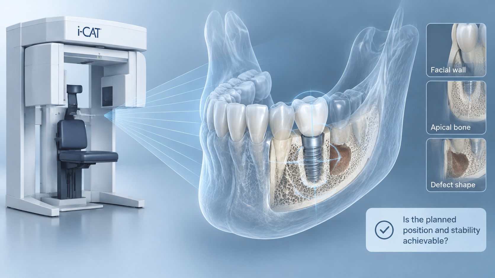
i-CAT 3D cone beam imaging helps the clinician assess the facial wall, available bone beyond the socket, and the shape of bone defects in three dimensions. The practical question is whether the intended implant position has enough support, rather than whether bone is present somewhere nearby.
X-Nav dynamic surgical navigation guides implant positioning during surgery against the planned location. Imaging helps establish the plan; navigation helps the surgeon follow it. Neither replaces the biological healing assessment.
For an anxious patient, this distinction can make the process easier to understand. The scan answers questions about anatomy, while follow-up examinations help determine whether the tissues are ready for the next procedure.
Different grafts answer different problems
Bone grafting rebuilds jawbone after tooth loss so that implants can be placed. The procedure chosen depends on what is missing:
- Ridge preservation helps maintain bone width and height after extraction.
- Ridge augmentation rebuilds a deficient ridge when the existing bone cannot support the planned treatment.
- Sinus augmentation increases available bone in the upper back-tooth region.
A barrier membrane may help protect a healing defect and support regeneration when socket walls are damaged or the surrounding gap needs additional support. The explanation of membranes after tooth extraction describes why that material may be included in a grafting plan.
A small socket-preservation procedure and reconstruction of a substantial bone defect shouldn't carry an identical timetable. The periodontist needs to assess what the graft accomplished and whether the developing site can provide primary stability.
A staged plan with reassessment and 3D imaging is often more predictable than promising a standard implant date.
Grafting therefore adds a healing requirement, not an automatic reset to a universal starting point. When thin gums or periodontal inflammation are also present, bone and soft tissue treatment need to be coordinated rather than scheduled independently.
Gum Disease and Recession Before Implant Surgery
Active periodontal disease can affect the surgical site and the long-term treatment plan. Replacing a tooth doesn't remove the need to manage inflammation elsewhere in the mouth.
Treatment depends on the diagnosis. Scaling and root planing cleans affected root surfaces below the gumline, followed by periodontal maintenance to monitor and manage the disease.
Selecting gum treatment
Surgical options may include LANAP, the Laser-Assisted New Attachment Procedure, using the FDA-cleared PerioLase MVP-7 laser, or traditional osseous surgery when clinically indicated. Laser treatment uses laser energy to address periodontal disease rather than making every patient follow the same surgical approach.
Many LANAP patients report less discomfort and a quicker return to routine activities than with traditional gum surgery. Most return to normal activity within 24 hours, although individual recovery and the timing of later implant treatment vary.
Gum recession requires a separate assessment. Soft tissue grafting treats recession and exposed tooth roots and can increase the firm, keratinized tissue around a site. When tissue is thin, grafting may be appropriate before implant placement; the options for gum recession treatment depend on the location and tissue needs.
A coordinated treatment sequence
- Control infection and inflammation. Active disease needs management before implant surgery so the clinician can evaluate a more stable tissue environment.
- Reassess the gums and bone. Examination and appropriate imaging help determine what support remains after treatment.
- Graft soft tissue when needed. The goal is to address a specific tissue deficiency, not to add a procedure automatically.
- Place the implant on the planned timeline. The date follows the response to treatment and the readiness of the site.
For patients worried about gum surgery, a clear sequence can reduce uncertainty. The consultation should distinguish which procedures are necessary, which are conditional, and how healing will be evaluated before another procedure is scheduled.
Individual results vary. A periodontist can determine whether disease control, gum grafting, bone treatment, or a combination should come before implant placement.
Book a Timing Consultation at Our Nearest Office
A personal implant timeline comes from an examination, 3D imaging, and a review of infection control, bone support, and gum health. The consultation establishes which option is realistic and what must happen before a date is set.
Periodontal Associates of Delaware is a board-certified periodontal practice with offices in Wilmington, Newark, and Dover. Its periodontists hold diplomate status with the American Board of Periodontology, and no referral is needed to schedule.
Preparing for the visit
- Bring available records: Recent X-rays, extraction notes, and previous grafting records clarify care already completed.
- List medications and health conditions: Include diabetes and smoking so the clinician can assess healing.
- Discuss the timing option: Ask which approach fits the extraction site and why.
- Clarify possible changes: Ask how bone grafting or gum treatment would affect the proposed sequence.
Whichever office you choose, the first visit is about understanding the site, not committing to a date. Expect a defined next step: what gets measured, what gets watched during healing, and which findings would move surgery earlier or later.
A useful plan names the next step and the findings needed to proceed. That clarity matters more than a calendar estimate, since the site's healing response and any grafting decide when surgery can follow.
Periodontal Associates of Delaware offers periodontal evaluation and implant planning to determine whether a site is ready for placement or needs healing or grafting first. Visit Periodontal Associates of Delaware to learn about care, then request an appointment at the nearest Wilmington, Newark, or Dover office for a timing consultation.