Gum recession is far more common than many adults realize. A 2024 systematic review and meta-analysis combining 15 studies and 37,460 participants estimated a 78.16% pooled global prevalence using the studies' minimum reported thresholds. The populations and examination methods differed substantially, so that figure isn't a prediction for any individual, but it makes an important point: receding gums aren't an unusual cosmetic concern.
Gum recession treatment starts with the cause and clinical measurements, not the size of the visible gap. Some adults need help controlling inflammation or changing brushing habits. Others benefit from surgery that covers exposed roots, thickens vulnerable tissue, or both.
Table of Contents
- Treatment Options for Gum Recession at a Glance
- What Causes Gum Recession and Why It Matters
- How a Periodontist Diagnoses Gum Recession
- Non-Surgical Care and Monitoring for Stable Recession
- How Gum Grafts and Flap Procedures Work
- Substitute Materials and Minimally Invasive Options
- Results, Recovery and Realistic Expectations
- Book a Gum Recession Evaluation Near You
Treatment Options for Gum Recession at a Glance
Several treatments can manage recession, and surgery isn't always the first step. The right choice reflects the depth of the defect, gum thickness, supporting tissue, and the reason the gum margin moved.
The American Academy of Periodontology's surgical procedures guide explains that graft surgery uses tissue from the palate or another donor source to cover an exposed root. Its purposes include helping prevent additional recession and bone loss, protecting vulnerable roots, and potentially reducing sensitivity.
Gum recession treatment options compared
| Option | Typically suited for | What it does | Main trade-off |
|---|---|---|---|
| Monitoring and non-surgical care | Shallow, stable recession that isn't progressing | Controls plaque, changes brushing, and manages sensitivity | Recession can continue if the cause remains |
| Coronally advanced flap (CAF) | Isolated defects with healthy surrounding tissue | Moves existing gum tissue over the exposed root | Less predictable with thin tissue or interdental bone loss |
| Tunnel technique | Multiple or adjacent recession defects | Creates tunnels beneath the gum through which tissue can be positioned | Results depend on tissue thickness and defect pattern |
| Connective tissue graft (CTG) | Thin tissue, limited keratinized gum, or deeper defects | Adds the patient's own palatal tissue beneath the gum | Creates a palate donor site that can cause discomfort |
| CAF plus CTG | Single defects needing thicker tissue and improved appearance | Combines repositioned gum with added tissue | More surgical work than a flap alone |
| Substitute materials | Patients hoping to avoid a palate donor site | Uses a processed tissue or collagen matrix without harvesting palatal tissue | Coverage may be lower than with the patient's own graft in some studies |
| Pinhole and laser-assisted options | Selected cases suitable for small entry points or an adjunctive approach | Repositions tissue through small openings, or uses laser energy as an adjunct | Not every defect qualifies, and supporting evidence differs |
A substitute isn't necessarily “donor-free.” Some materials come from processed donated tissue, while others come from animal-derived collagen. The important distinction for many patients is whether tissue must be taken from their own palate.
Practical rule: The table is a starting map, not a way to select surgery from a photograph.
Patients can identify the rows that resemble their situation and bring questions to a consultation. A visible gap alone doesn't reveal whether enough firm gum remains, whether bone support has changed, or whether inflammation needs treatment first.
What Causes Gum Recession and Why It Matters
A systematic review and meta-analysis estimated that 81.1% of people have recession of at least 1 mm, while 16.2% have recession of at least 5 mm. The researchers also reported associations between recession and several factors. An odds ratio describes an association, not proof of what caused recession in an individual.
Recession contributors and what the exam checks
| Contributor | Reported association or finding | What the exam checks |
|---|---|---|
| Periodontitis | Strongest reported association, odds ratio 9.90 | Probing depths, bleeding, attachment levels, and bone levels |
| High frenum | Odds ratio 4.58 | Whether the band connecting the lip or cheek to the gum pulls on the margin |
| Dental plaque | Odds ratio 4.26; the regional study reported it as a contributor in 44.1% of its population | Plaque deposits and inflammation along the gumline |
| Occlusal trauma | Odds ratio 3.20 | Bite forces, tooth position, mobility, and wear patterns |
| Faulty or aggressive brushing | The regional study reported it in 42.7% of its population | Brushing pressure, technique, and tooth-surface wear |
| Tobacco use | A concern for healing and recession progression | Smoking history and current use |
The regional findings for plaque and brushing come from a different study population than the pooled associations. Percentages describe groups, not the cause of a particular person's recession.
Age, brushing pressure, tissue pull, inflammation, and bite forces are among the factors a clinician considers when examining recession. The connection between teeth grinding and receding gums also calls for assessment rather than an assumption based on tooth wear alone.
Covering an exposed root may improve protection, but it does not necessarily address the contributor that exposed it. Treatment options depend on the cause and the measurements, not only on how large the visible gap looks. Stable, shallow recession may be monitored, while progression, sensitivity, limited protective tissue, or appearance concerns may support treatment.
How a Periodontist Diagnoses Gum Recession
Diagnosis comes before choosing a procedure. Measurements distinguish a root-coverage problem from active periodontal disease and show whether existing tissue can be repositioned or needs reinforcement.
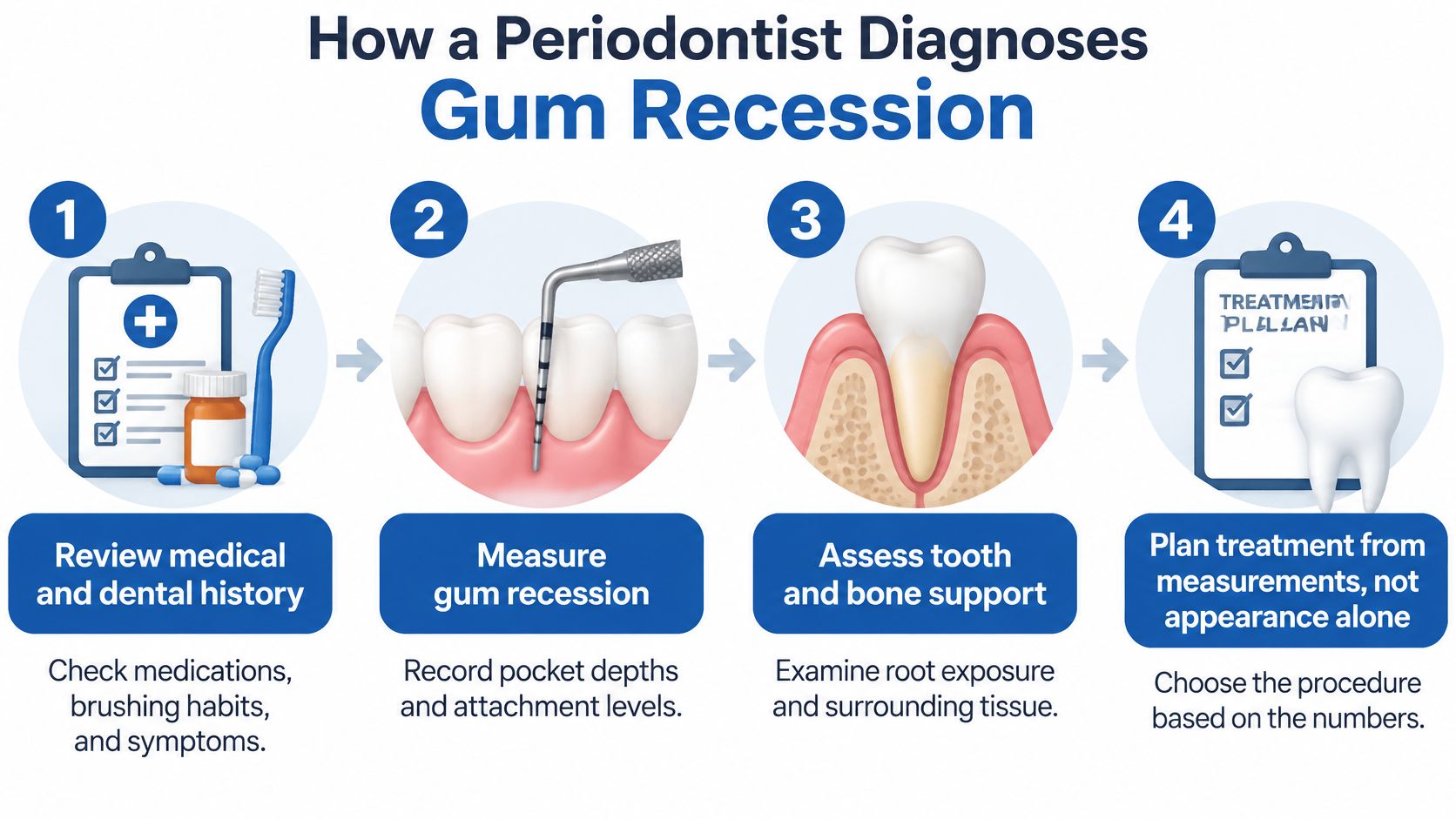
The measurements behind the treatment plan
Review medical and dental history. Medications, smoking, grinding, brushing habits, and previous gum treatment can affect planning. Symptoms and older photographs may help establish when changes began.
Check plaque and inflammation. The clinician examines bleeding and measures periodontal pockets. Active inflammation needs control before elective root-coverage surgery.
Measure recession depth. The reference point is the cemento-enamel junction, where the tooth's crown meets its root. Measuring from that landmark to the gum margin provides a repeatable record rather than a visual estimate.
Measure keratinized tissue width. This is the firmer band of gum extending below the recession toward the movable lining of the mouth. Clinical guidance on recession treatment identifies less than about 2 mm of this tissue and gum thickness below approximately 1 mm as findings that can change the surgical choice, often favoring added connective tissue.
Assess gum thickness. A thin covering over a prominent root presents a different challenge from a thick, resilient gum margin. The clinician evaluates whether adding volume would improve protection and stability.
Check attachment between teeth. Interproximal attachment loss means that support between neighboring teeth has been lost. That loss can limit how far a flap can predictably cover a root.
Inspect the exposed root. Cervical notches, fillings, and sensitivity may require coordination between periodontal and restorative care. A worn root surface can also make the original crown-root boundary harder to identify.
Evaluate bone when indicated. Conventional dental imaging may answer the initial questions; additional imaging isn't automatic. When closer three-dimensional evaluation is justified, i-CAT 3D cone beam imaging can help assess bone anatomy.
Understanding the defect classification
The Miller classification describes recession in relation to surrounding tissues and support. Class I and II defects generally retain more favorable support between teeth, while Class III defects have features that make complete coverage less predictable.
Periodontal Associates of Delaware uses i-CAT imaging when indicated and offers LANAP laser gum surgery with the FDA-cleared PerioLase MVP-7 for periodontal disease. These technologies serve different purposes; neither replaces the clinical measurements needed to plan recession treatment.
A useful written plan records the affected teeth, recession depth, tissue quality, attachment findings, and the reason for the recommended approach.
Non-Surgical Care and Monitoring for Stable Recession
Monitoring can be an appropriate treatment plan, not a sign that a concern has been ignored. It suits selected shallow defects when measurements remain stable and the contributing factors can be controlled.
Non-surgical care aims to protect the remaining tissue and improve comfort. It doesn't rebuild a missing gum margin.
A practical care plan
- Gentler brushing: A soft-bristle brush and light pressure reduce repeated trauma. A dental professional can demonstrate small, controlled movements instead of forceful horizontal scrubbing.
- Professional plaque control: Cleaning and periodontal maintenance address deposits that home care can't remove. Active periodontal disease may require scaling and root planing rather than a routine cleaning alone.
- Sensitivity management: Desensitizing toothpaste, fluoride products, or professionally applied treatments may make exposed roots more comfortable. Persistent or localized pain needs examination for other causes.
- Bite-related care: A nightguard may protect teeth when grinding is present. Bite adjustment shouldn't be automatic and requires a specific diagnosis.
- Tooth-position assessment: Orthodontic consultation may help when a tooth's position contributes to tissue vulnerability. Treatment must account for the surrounding bone and gum support.
- Tobacco cessation: Stopping tobacco use supports periodontal health and healing, whether or not surgery becomes necessary.
A stable gum margin can be a worthwhile outcome even when the exposed root remains visible.
The periodontist sets the monitoring interval according to disease activity and risk. Comparing recorded measurements is more reliable than repeatedly checking the mirror, where lighting and viewing angles can make small differences look dramatic.
A concrete step this week is to note which foods trigger sensitivity, replace an overly firm toothbrush, and arrange a periodontal examination. Home photographs can supplement that record, but they can't establish whether bone or attachment has changed.
How Gum Grafts and Flap Procedures Work
Consider an adult with an exposed root and thin surrounding gum. Moving that thin tissue alone may not provide the thickness the clinician wants, so the plan may combine a flap with a connective tissue graft.
A flap repositions existing gum toward the tooth's crown. A graft adds tissue. These aren't competing procedures in every case; a periodontist may use them together.
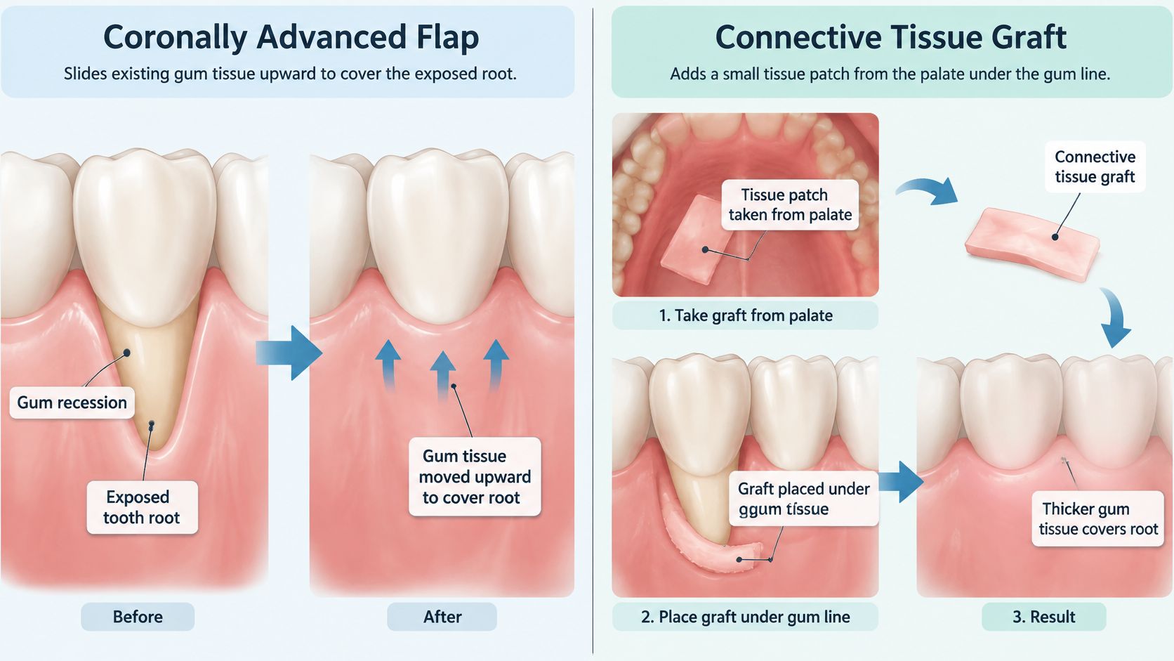
What the comparisons mean
A systematic review and meta-analysis comparing flap and tunnel procedures found no statistically significant difference in mean root coverage overall, with a reported mean difference of 2.93 percentage points. That doesn't establish that the techniques are interchangeable for every defect: isolated recession, adjacent defects, tissue thickness, and available support still guide selection.
A separate Cochrane review of root-coverage procedures found that connective tissue grafting with a coronally advanced flap produced slightly greater recession reduction than a resorbable guided tissue regeneration membrane with a flap, by 0.37 millimeters. A modest average difference can inform planning, but it doesn't identify the best procedure without an examination.
A typical connective tissue graft appointment
The treatment area is numbed. Local anesthesia is administered around the recipient site and palate. The clinician checks anesthesia before proceeding; patients should report sharp sensation rather than assuming it must be tolerated.
The gum around the recession is prepared. The periodontist creates space for the graft using the selected flap or tunnel approach. Root-surface preparation may also be needed.
Connective tissue is collected from the palate. The clinician takes a suitable tissue sample from beneath the palatal surface. The amount and shape reflect the recipient site's needs.
The graft is positioned and secured. Sutures stabilize the graft and surrounding gum. Keeping the tissue still during early healing helps protect the surgical repair.
Both sites receive a protection plan. Instructions explain cleaning, eating, prescribed products, and follow-up care. A protective appliance or dressing may be used when appropriate.
The palate donor site is often a major source of worry. It can cause soreness, and discomfort differs among patients; the care plan should explain how the site will be protected and which pain-control measures are appropriate.
Before treatment, the clinician should also clarify the main objective. Increasing tissue thickness, improving root coverage, and improving gumline appearance are related goals, but they aren't identical, and a procedure may prioritize one over another.
Substitute Materials and Minimally Invasive Options
A palate donor site can be avoided with substitute materials, but the choice depends on the defect's measurements and treatment goals. The size of the visible gap alone does not determine which option fits.
A systematic review of substitute materials reported greater mean root coverage with connective tissue grafts than with xenogeneic collagen matrices and porcine acellular dermal matrices at 6 and 12 months. A 2025 review of acellular dermal matrix for recession defects found that most included studies judged it comparable to connective tissue grafting for Miller Class I and II defects, while several favored the patient's own tissue.
Reviews can reach different conclusions because they examine different materials, procedures, patient groups, and outcomes. Substitute materials may also help when treatment involves numerous sites and the available palatal tissue is insufficient.
Choosing the trade-off
- Defect type and support: Recession pattern and attachment between the teeth affect the potential for coverage.
- Tissue thickness: The material must provide the volume needed for the planned repair.
- Number of teeth: The number of sites affects how much graft material is needed.
- Donor-site preference: The patient's willingness to have palatal surgery is part of the decision.
- Appearance and retreatment: The plan should account for likely contour, coverage, and the possibility of further treatment.
The Pinhole Surgical Technique uses small entry points to loosen and reposition gum tissue through a tunneling approach. Suitability and long-term predictability depend on the individual defect.
LANAP treats periodontal disease. It does not replace a gum graft for recession. The role of laser adjuncts in recession treatment remains under study.
A discussion of gum graft alternatives should address the measured defect and compare the likely coverage, appearance, donor-site considerations, and potential need for further treatment.
Results, Recovery and Realistic Expectations
Root coverage can be substantial, but complete coverage isn't guaranteed. Mean coverage describes how much of the exposed root was covered on average; complete coverage describes how often no recession remained at the measured site.
Reported root coverage outcomes
The periodontal evidence summary on root coverage provides the average outcomes and success definitions below. The trial ranges come from the Cochrane review's randomized-trial results.
| Measure | Reported result | Context |
|---|---|---|
| Mean root coverage, Miller Class I and II | About 70% | Average across systematic review data |
| Complete root coverage, Miller Class I and II | About 35% | Results differ by defect and patient |
| Success threshold, Miller Class I and II | At least 70% coverage | Definition used in the evidence summary |
| Success threshold, Miller Class III | At least 50% coverage | Definition for more advanced defects |
| Complete coverage, flap with graft versus flap alone | 53% versus 28% | Single recession defects in a systematic review |
| Complete coverage, connective tissue graft trials | 18.1% to 95.6% | Randomized trials of single defects |
| Complete coverage, coronally advanced flap trials | 7.7% to 81.8% | Randomized trials of single defects |
Success thresholds are study definitions, not promises or universal standards. The flap-plus-graft comparison supports adding tissue in suitable cases, but it doesn't establish the same advantage for every patient.
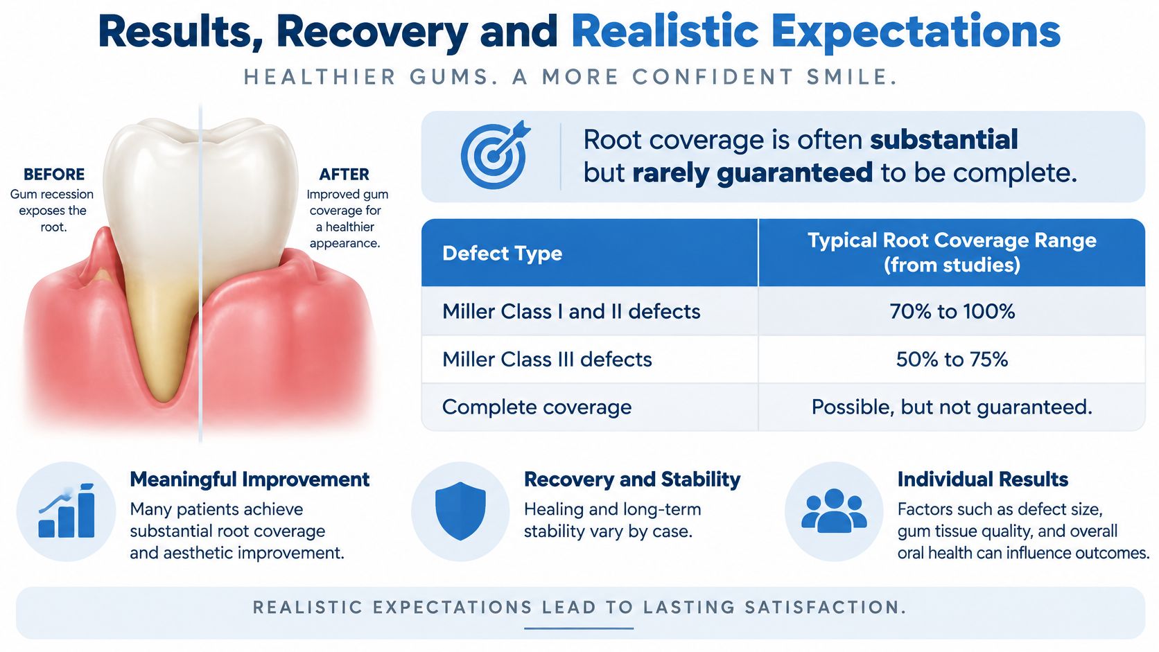
Published ranges are broad because recession depth, tissue thickness, root anatomy, plaque control, smoking, and healing differ. The measured averages and study definitions in the table are more useful than treating any broad visual range as a personal forecast.
Recovery and protecting the repair
Early recovery may include swelling, soreness, and a soft diet. Sutures are checked and removed or allowed to dissolve according to their type and the surgeon's plan; gum surgery stitch care should follow the procedure-specific instructions.
Healing continues after initial tenderness improves, and gum color and contour can take longer to settle. Increasing pain, persistent bleeding, or concerns about graft movement warrant contact with the treating team.
- Clean as instructed: Brushing directly over a new graft may be restricted initially.
- Avoid tobacco: Healing tissue needs protection from tobacco exposure.
- Resume gentle technique: Regular brushing should follow clearance and demonstration.
- Attend follow-ups: The clinician checks healing before judging the final result.
Individual results vary. A periodontist can estimate likely outcomes only after evaluating the specific defect and health history.
Book a Gum Recession Evaluation Near You
Sensitivity to cold or sweets, teeth that look longer, or a gumline that appears to be receding are reasons to arrange an evaluation. Bleeding gums or loose teeth also merit attention, since recession can occur alongside periodontal disease.
An examination helps determine whether monitoring, non-surgical care, or a root-coverage procedure fits the cause and tissue measurements. You can ask questions and discuss options before making a treatment decision.
Arranging a consultation
Periodontal Associates of Delaware serves Wilmington, Newark, Dover, Rehoboth Beach, Pike Creek, and Hockessin, Delaware. Its periodontists hold diplomate status with the American Board of Periodontology. You do not need a referral to schedule.
- Choose a location: The practice has offices in Wilmington, Newark, Dover, and Rehoboth Beach.
- Request an appointment: Use the online appointment request form or call (302) 995-6979.
- Bring useful information: Note your symptoms, medications, and any previous gum treatment.
Ask which procedure is recommended and why it fits your measurements. You can also ask about anesthesia, care for a palate donor site, and the expected costs. Most major insurance plans are accepted, and flexible financing is available. Confirm your specific coverage with the practice.
Periodontal Associates of Delaware provides periodontal evaluation and gum grafting for exposed roots. Patients can request an appointment to discuss the cause of recession and suitable care options.