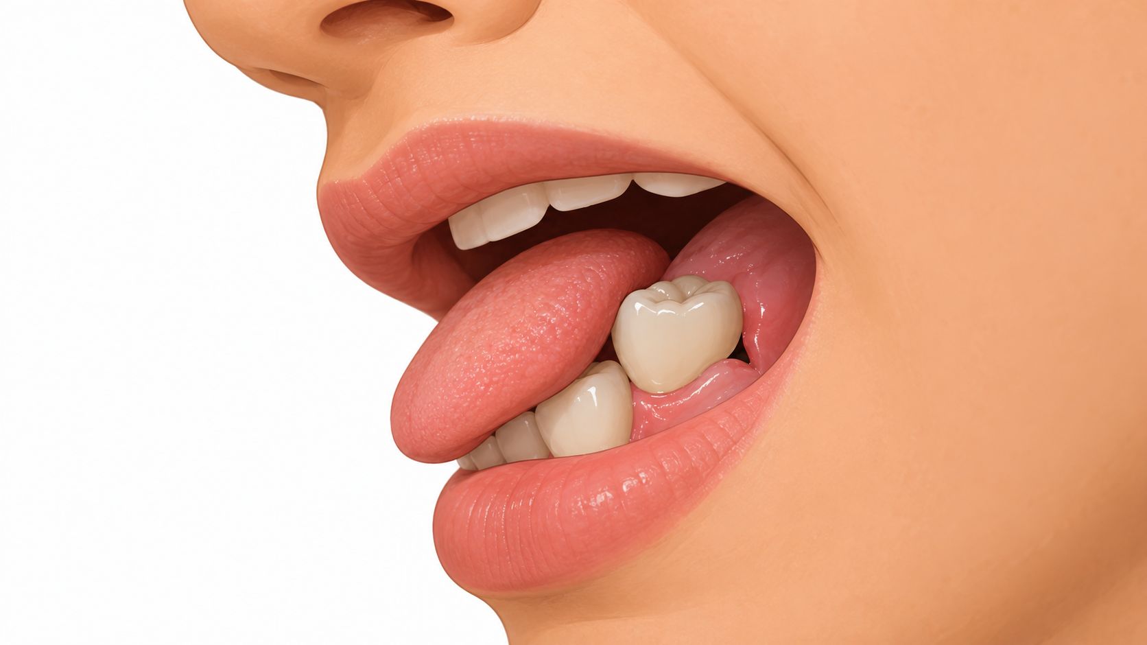
A tongue catches on a small ledge beside a dental crown. In the mirror, a dark line appears where the gum once covered the edge. For an adult noticing gums receding around a crown, the immediate worry may be a failing restoration, a damaged tooth, or gum surgery. The visible change deserves attention, but it doesn't reveal the cause by itself.
Table of Contents
- Noticing the Gumline Pulling Back From a Crown
- What Gum Recession Around a Crown Actually Means
- The Five Causes Clinicians Actually Look For
- What a Periodontal Evaluation Looks Like Step by Step
- Treatment Options From Watchful Waiting to Grafting
- When the Crown and the Gums Need Coordinated Care
- Daily Habits That Protect the Gumline Around a Crown
- Your Next Step and How to Get Evaluated Locally
Noticing the Gumline Pulling Back From a Crown
A crown that once blended into the smile may suddenly look longer. Someone might first notice the change while flossing, comparing photographs, or feeling sensitivity during a cold drink. Even a small difference can feel alarming when the tooth has already needed restorative treatment.
An exposed crown edge doesn't automatically mean the crown has failed. The gum may have moved because of inflammation, tissue anatomy, brushing trauma, or a problem with the restoration. Sometimes several factors overlap, which is why a photograph alone can't settle the question.
Common doesn't mean harmless
Gum recession is widespread among adults, not something that happens only around crowns. A systematic review covering 22 studies estimated that 81.1% of people had at least one site with recession of 1 mm or more, although that finding doesn't identify the cause of recession in any particular patient (systematic review of gingival recession prevalence).
A newly noticed margin, by itself, isn't proof of an emergency or a need for surgery. It is a reason for a calm examination, especially when the area has changed or become difficult to clean. Treatable problems can look similar at the surface while requiring different kinds of care.
A useful starting point: The question isn't only whether the crown caused the recession. It's whether the crown is revealing a problem in the surrounding gums, supporting tissues, or tooth position.
Evaluation comes before a surgical decision
Fear of gum surgery can lead an adult to postpone even an examination. Yet an assessment may lead to monitoring, changes in home care, periodontal treatment, crown correction, or grafting, depending on the findings.
Persistent bleeding, pus, increasing tooth mobility, or a visibly advancing gumline deserves prompt professional assessment. The important distinction is between noticing a change and knowing why it happened. A periodontist gathers that information before recommending a procedure.
What Gum Recession Around a Crown Actually Means
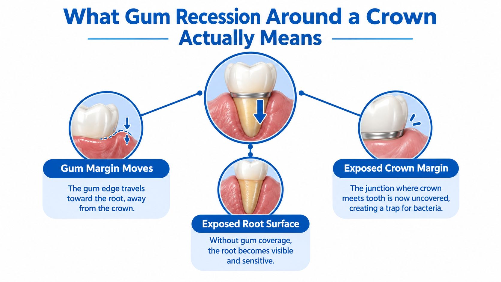
Gum recession means the edge of the gum has moved toward the tooth's root. Around a crowned tooth, that movement can uncover the natural root, the edge of the crown, or both. The crown margin is the junction where the restoration ends against the tooth.
A useful analogy is a picture frame that slips down a wall and leaves part of the artwork uncovered. The newly visible area doesn't necessarily mean the artwork moved or broke. Similarly, an exposed crown margin may reflect movement of the surrounding tissue rather than movement of the crown.
The landmark beneath the restoration
On a natural tooth, clinicians use the cemento-enamel junction, or CEJ, as a reference point. This is the boundary between the enamel covering the tooth's upper portion and the cementum covering its root. Recession is measured in millimeters from that boundary to the gum edge, rather than estimated from how long the tooth appears (clinical measurement of gingival recession).
A crown or filling may hide the CEJ. In that situation, the periodontist may need another stable landmark and additional clinical assessment. Comparing visible crown height alone can give a misleading impression of how much tissue has changed.
- A longer-looking tooth describes an appearance, not a complete diagnosis.
- An uncovered crown margin identifies a visible restorative boundary.
- Attachment loss concerns the tissues securing the tooth and requires periodontal assessment.
Why the distinction matters
A crown restores tooth structure, but it doesn't replace lost gum attachment or supporting bone. Making a new crown longer could change the appearance without addressing the underlying tissue problem.
An exposed root may also be sensitive or require protection from decay. An uncovered margin may remain smooth and cleansable, or it may contain an irregularity that holds plaque. Those differences matter more clinically than the darkness or size of a line seen in the mirror.
The Five Causes Clinicians Actually Look For
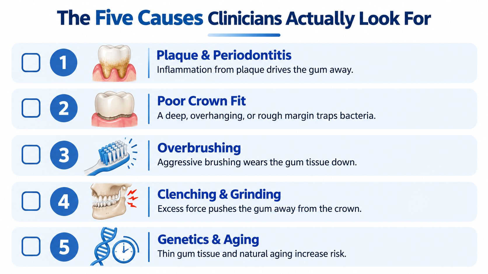
Clinicians generally assess inflammation and crown cleanability early, because either can require treatment before cosmetic correction makes sense. That is a practical diagnostic priority, not a claim that either cause explains every case. Gum anatomy, daily habits, and tooth position also deserve attention.
Inflammation and the restoration
Plaque-driven inflammation or periodontitis can affect a crowned tooth just as it affects other teeth. Bleeding during brushing may be a clue, but it doesn't establish how much supporting tissue has been lost. Periodontitis involves the tooth's supporting structures, not just a red gum edge.
A deep, overhanging, or rough crown margin may create an area that is difficult to clean. A visible ledge or persistent irritation can raise suspicion, although the patient may not be able to detect the defect. Historical research linked deeper restorative margins with later tissue changes, making margin position an important part of the examination (restorative margin position and periodontal recession).
Tissue, technique, and tooth position
The remaining possibilities require a different kind of investigation:
- Thin gum tissue. A thin periodontal phenotype means the gum has less thickness. Tissue may look delicate or nearly translucent, but a clinician must assess it rather than rely on appearance.
- Traumatic brushing or flossing. Forceful horizontal scrubbing or repeatedly snapping floss against the gum can contribute to injury. Sensitivity near a receded area may bring attention to the problem without proving its cause.
- Tooth position and supporting anatomy. A tooth positioned toward the outer edge of its bone may have vulnerable tissue coverage. Bone dehiscence means part of the bony covering over the root is absent.
Bite forces belong in the assessment, but clenching or grinding shouldn't automatically receive the blame. The relationship between grinding teeth and receding gums needs to be interpreted alongside inflammation, tissue thickness, and tooth position.
A garden edge offers a gentle comparison: trouble may develop where the soil is thin, the border is jagged, the watering is too forceful, or a plant leans outward. Likewise, the gumline can change for different reasons. Age or inherited tissue characteristics shouldn't become shortcuts that replace a proper diagnosis.
What a Periodontal Evaluation Looks Like Step by Step
A periodontal consultation is an information-gathering visit, not consent to surgery. The periodontist examines how the crown, gum, root, and supporting bone relate to one another. For an anxious adult, understanding what each part of the examination answers can make the visit less mysterious.
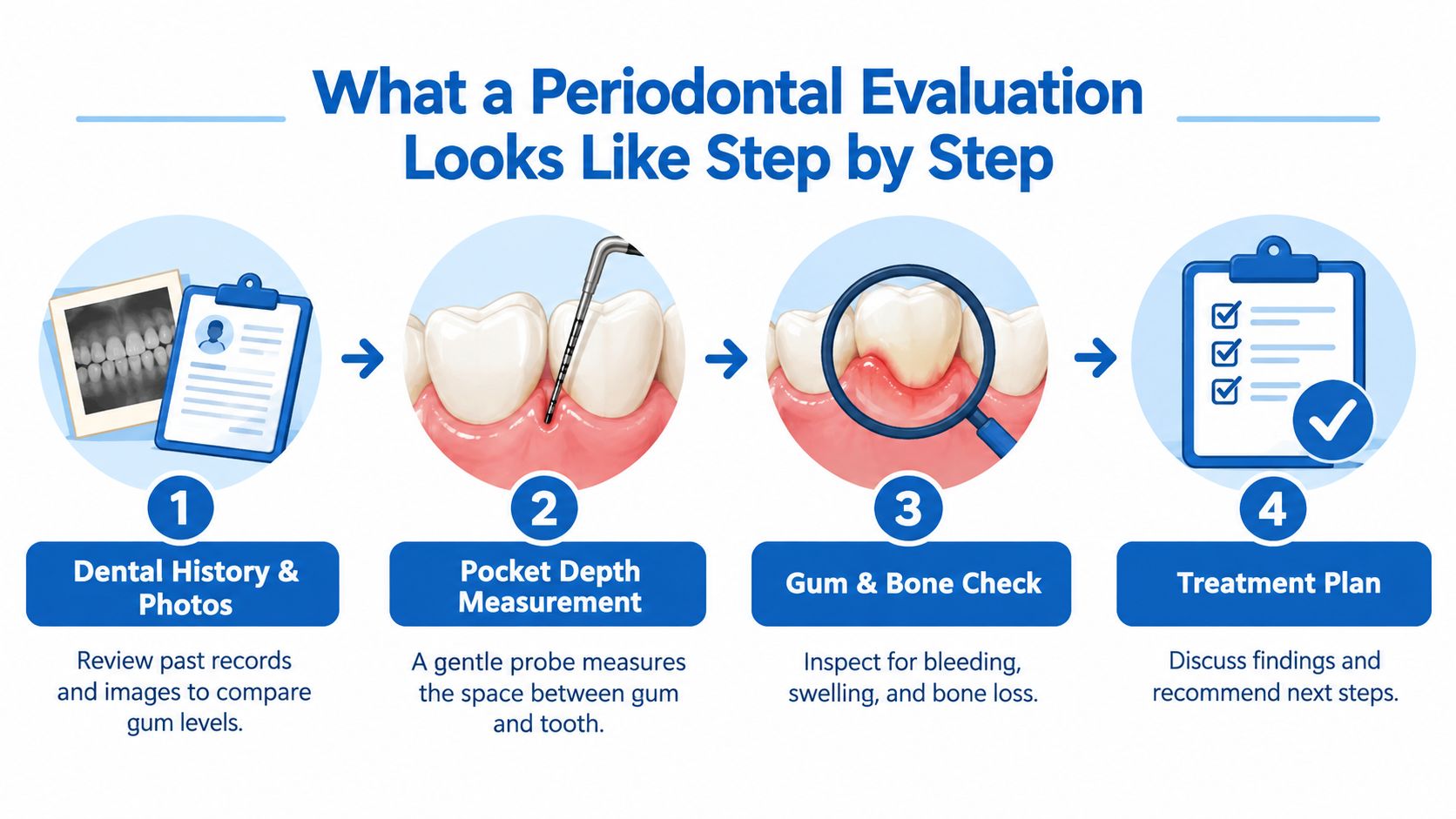
Establishing what changed
Older dental records and photographs can help distinguish a longstanding exposed edge from an advancing change. The history also includes when the crown was placed, whether symptoms began afterward, and whether other teeth show recession.
The clinical examination then addresses several questions:
- How deep is the space beside the tooth? A small periodontal probe measures the space between tooth and gum. Pocketing in teeth is different from visible recession, so clinicians record both.
- Does the tissue bleed when assessed? Bleeding provides information about inflammation. The periodontist interprets it with the other findings rather than as a diagnosis by itself.
- How much protective tissue remains? Tissue thickness and the width of firm, keratinized gum help guide treatment selection.
- Can the crown margin be maintained? The examination checks contour, overhangs, residual cement, and access for cleaning.
Looking beneath the gumline
Bitewing or periapical dental images may help assess bone support and the relationship between the tooth and restoration. Three-dimensional imaging is selected when the clinical question calls for it, rather than being necessary for every exposed crown edge.
The practice's i-CAT 3D cone beam imaging can provide additional information about bone and root position when indicated. Imaging supports the examination; it doesn't replace direct assessment of gum inflammation or crown cleanability.
These findings form part of a full periodontal evaluation that includes probing depths, attachment levels, recession dimensions, crown contour, and supporting bone (clinical assessment of restorative-periodontal relationships). Persistent sensitivity, suppuration, or increasing mobility also informs the treatment plan.
An effective consultation explains the likely cause, the amount and pattern of tissue loss, and whether the crown is contributing to the problem or simply making it visible.
Patients should also hear what remains uncertain. When earlier records aren't available, follow-up measurements may help establish whether a noninflamed area is stable. A careful explanation is more useful than an immediate recommendation based only on the visible gap.
Treatment Options From Watchful Waiting to Grafting
Treatment follows the diagnosis, not the appearance of the crown edge. A stable, cleansable area may need a different approach from a bleeding site with a defective restoration. The amount of exposed root matters, but so do symptoms, remaining attachment, tissue thickness, and the patient's priorities.
Matching Treatment to the Cause of Recession Around a Crown
| Likely Cause | First-Line Treatment | When Grafting Is Added |
|---|---|---|
| Stable, noninflamed recession around a maintainable crown | Observation, tailored hygiene, and periodontal maintenance | When tissue protection, sensitivity, or appearance warrants it |
| Plaque-related inflammation or periodontitis | Control inflammation and treat periodontal disease | After disease control, if a suitable tissue defect remains |
| Defective crown margin or contour | Correct or replace the restoration when indicated | When correction alone won't address the tissue deficiency |
| Thin tissue with exposed root | Reduce trauma and assess protective tissue needs | When added thickness or root coverage is appropriate |
| A defect involving both crown and root | Coordinated restorative-periodontal planning | As part of a combined plan when anatomy permits |
Observation is active monitoring, not neglect. It means documenting the area, making it easier to maintain, and reassessing it. A well-fitting crown may not need replacement when recession is stable, noninflammatory, and cleansable (gum recession assessment and management).
A defective restoration calls for a different response. Depending on its fit and contour, correction or replacement may be appropriate. Recession alone, however, isn't enough evidence to remove a crown.
What grafting may accomplish
Soft-tissue grafting may add gum thickness, cover an exposed root, and improve protection or sensitivity. Depending on the procedure and clinical circumstances, options may include the patient's own tissue or donor material. The material and technique require an individualized discussion.
In a clinical study of 32 crowned teeth with suitable recession defects, connective-tissue grafting achieved 85.1% mean root coverage; an evidence review also reported approximately 80% root coverage for a coronally advanced flap combined with a graft (root-coverage approaches and outcomes around crowned teeth). These are study averages, not promises for an individual tooth.
A coronally advanced flap repositions gum tissue toward the crown, while a connective-tissue graft adds tissue beneath it. Tunneling approaches may also be considered for suitable defects. Available attachment and the crown-to-root transition influence what coverage is achievable.
Fear of grafting deserves a direct conversation about the proposed technique, recovery, alternatives, and realistic objective. A discussion of gum graft failure rates and factors affecting healing can support informed questions without turning a population average into a personal forecast.
A graft isn't a substitute for disease control or correction of a plaque-retaining crown. Sometimes improved thickness and comfort are more appropriate goals than complete coverage of every visible margin.
When the Crown and the Gums Need Coordinated Care
Replacing a crown and treating the gum aren't always separate decisions. A defect near the gumline may extend across both the natural root and the restoration. In that situation, the restorative dentist and periodontist need to agree on the final tooth contour and the tissue position that can reasonably be achieved.
Planning the boundary between tooth and gum
The biologic soft-tissue attachment is the protective attachment surrounding the tooth near the bone. A crown margin placed into this space can interfere with tissue health. Its position must be considered along with fit, contour, and the patient's ability to clean it.
Persistent inflammation around a crown margin, bleeding on probing, pocket formation, and attachment loss are recognized clinical signs that the restoration may be impinging on the biologic soft-tissue attachment (clinical signs of restorative attachment interference). These findings require investigation rather than a cosmetic attempt to hide the edge.
When a cervical defect crosses root and crown surfaces and the natural reference boundary can't be identified, combined restorative-periodontal treatment may be indicated (guidance on combined restorative-periodontal care). The plan may involve correcting a margin, rebuilding tissue, or staging both.
Sequencing matters: A definitive crown should be planned around a maintainable, assessed tissue position rather than placed in the hope that the gum will later adapt.
Choosing technology for the actual problem
When active periodontal disease is present, LANAP laser gum surgery, using the FDA-cleared PerioLase MVP-7, may be considered within the broader treatment plan. LANAP addresses periodontal disease; it shouldn't be presented as a substitute for root-coverage grafting.
If separate findings indicate a need for tooth replacement, X-Nav dynamic surgical navigation may support implant planning and placement. Recession around a natural-tooth crown does not, by itself, establish a need for an implant.
Tissue stabilization may need to occur before the final restoration so the dentist can place its margin in a maintainable location. The sequence varies with the defect and disease status. Coordinated care helps avoid treating the visible crown edge while leaving the underlying tissue problem unresolved.
Daily Habits That Protect the Gumline Around a Crown
Daily care should remove plaque without repeatedly injuring the gum edge. More pressure isn't a better solution to bleeding or a rough-feeling margin. If an area remains difficult to clean despite careful technique, the crown's contour needs professional assessment.
Gentle cleaning with a clear purpose
A practical home-care approach includes the following:
- Use a soft-bristled brush. Angle the bristles toward the gumline and use gentle, controlled circular movements instead of aggressive horizontal scrubbing.
- Choose a nonabrasive toothpaste. Product selection should reflect exposed-root sensitivity and the dental team's recommendations.
- Guide floss carefully. Curve it against the tooth and move it along the surface rather than snapping it onto the gum.
- Match the interdental tool to the space. Floss, appropriately fitted interdental brushes, or a water flosser may be recommended according to access and tissue anatomy.
A hands-on demonstration can be more useful than general advice to brush better. The dental team can observe the patient's technique and identify where excess pressure or poor access creates difficulty.
Maintenance after treatment
Professional maintenance provides opportunities to reassess inflammation, cleanability, and tissue changes. The interval should reflect periodontal risk and treatment history rather than a universal schedule. Thin tissue and previous recession make an individualized maintenance plan particularly important.
Long-term research found that greater keratinized-tissue width after treatment was associated with better margin stability, while grafts using the patient's own tissue were associated with less subsequent recession increase (systematic review of long-term soft-tissue stability). That supports attention to tissue quality as well as appearance.
Brushing technique doesn't rebuild lost gum attachment. Its purpose is to protect the remaining tissue and support the conditions needed for treatment to last. After grafting, patients should follow procedure-specific cleaning instructions rather than immediately returning to their usual routine.
Your Next Step and How to Get Evaluated Locally
A changing gumline, bleeding, sensitivity, or a newly exposed dark margin calls for periodontal evaluation rather than an unexamined wait-and-see approach. The visible edge may be the concern that brings an adult to the appointment, but the treatment decision depends on the tissues around it.
A practical decision path
- Arrange an assessment when a crown's gumline changes or symptoms persist.
- Bring available records or older photographs that may help establish the history.
- Discuss the findings and alternatives before deciding on crown work or gum surgery.
Board-certified periodontal care is available through the practice's Wilmington, Newark, and Dover offices. These locations serve adults across New Castle and Kent Counties, including Pike Creek, Hockessin, Bear, Middletown, and surrounding communities, for gum disease, recession, bone loss, missing teeth, and implant-related concerns.
Anxiety about surgery can be discussed at the first visit. Individual results vary, and readers should consult a periodontist for diagnosis and treatment planning. An evaluation doesn't commit a patient to a procedure.
Periodontal Associates of Delaware offers periodontal evaluations, gum grafting, and coordinated restorative care for recession around crowns. Adults can request an appointment at the nearest Wilmington, Newark, or Dover office to learn whether observation, periodontal treatment, crown correction, or grafting may fit their situation. The first visit is an opportunity to understand the findings and discuss concerns before choosing treatment.