If bleeding gums stop after a cleaning, does that mean the disease is gone, or could damage remain below the surface? That question exposes an important gap in how gum disease is often understood: symptoms describe inflammation, but they don't reveal how much support a tooth has lost.
The difference between gingivitis and periodontitis comes down to the tissues involved, the measurements found during an examination, and the care needed afterward. Understanding that distinction can help adults approach a periodontal evaluation with less uncertainty, especially when surgery or tooth loss is a concern.
Table of Contents
- Gingivitis and Periodontitis Side by Side
- What Happens Below the Gumline
- How Dentists Tell the Two Apart
- Causes, Risk Factors and How Common Each Is
- Can Gingivitis Turn Into Periodontitis
- Treatment Options for Each Stage
- Book a Periodontal Evaluation If Warning Signs Appear
Gingivitis and Periodontitis Side by Side
Gingivitis is inflammation limited to the gum tissue, usually triggered by bacterial plaque; periodontitis is the destructive form in which inflammation extends below the gumline and damages the structures that hold teeth in place. Both can involve redness, swelling, tenderness, and bleeding during brushing or flossing. Their symptoms overlap, but their effects on tooth support are different.
The National Library of Medicine's clinical overview of gingivitis explains that gingivitis leaves the periodontal ligament, connective-tissue attachment, and alveolar bone intact. Those intact structures are why plaque removal and professional cleaning can generally reverse gingivitis, while periodontitis requires management of established support loss.
Gingivitis and periodontitis compared
| Feature | Gingivitis | Periodontitis |
|---|---|---|
| Tissue affected | Gum tissue only | Gum tissue, periodontal ligament and alveolar bone |
| Attachment to the tooth | Intact | Lost, measured as clinical attachment loss |
| Bone around the tooth | Intact | Lost as the disease advances, visible on X-rays |
| Common signs | Redness, swelling, tenderness and bleeding when brushing or flossing | Bleeding and inflammation, sometimes with little or no pain |
| Reversibility | Generally reversible once plaque is removed | Lost tissue is considered irreversible; infection can be controlled and progression slowed |
The periodontal ligament is the network of fibers connecting a tooth's root to the surrounding bone. The alveolar bone is the part of the jaw that supports the teeth. A tooth may still look normal above the gumline even when these deeper structures have been damaged.
A useful analogy is a railing supported by wooden posts. Gingivitis resembles surface rust on the metal railing that cleaning can remove before the structure is compromised. Periodontitis resembles rot in the post the railing is bolted to: cleaning the visible surface doesn't restore the weakened support underneath.
The analogy has limits. Periodontal treatment can sometimes rebuild selected areas through regenerative procedures, but that doesn't erase the history of disease or guarantee restoration of every lost structure.
Practical rule: The amount of blood in the sink doesn't measure the amount of bone supporting a tooth.
For someone noticing bleeding, the table provides context, not a home diagnosis. A periodontal examination is the reliable way to determine whether inflammation is limited to the gums or whether attachment and bone have been lost.
What Happens Below the Gumline
Healthy gum tissue fits closely around each tooth, leaving a shallow space along the tooth's surface. During daily cleaning, plaque removal helps keep this area free from a persistent buildup of bacteria. Problems begin when bacterial biofilm remains and the surrounding tissue becomes inflamed.
Consider an adult whose back teeth are difficult to clean. Plaque stays near the gumline, the tissue swells, and gentle brushing begins to cause bleeding. At this point, the inflammation may still be confined to the gums.
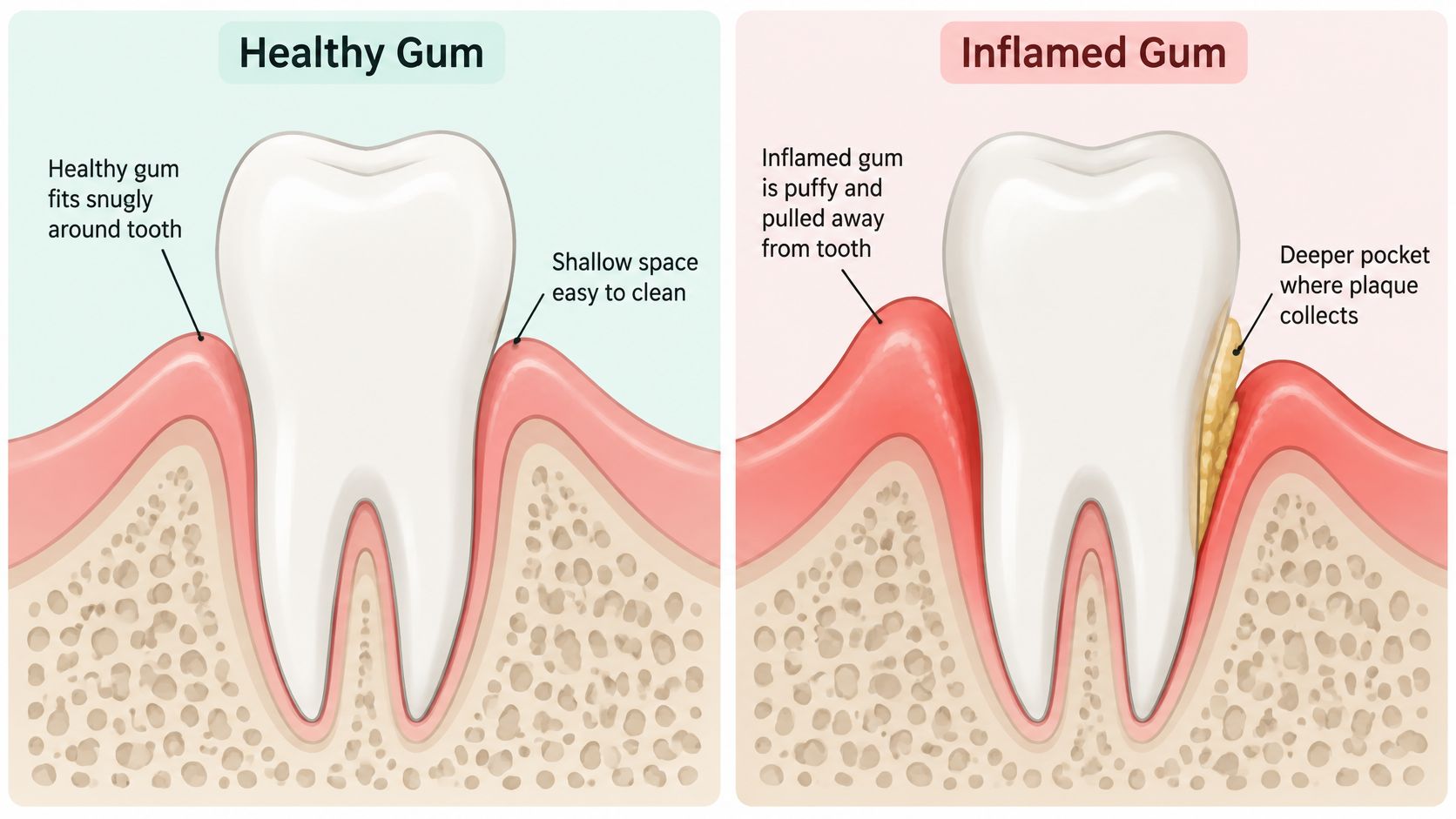
How inflammation can reach tooth support
If destructive inflammation develops, the attachment between the tooth and surrounding tissues breaks down. The space beside the root can deepen into a periodontal pocket, where bacteria become harder to remove with ordinary home care.
The National Institute of Dental and Craniofacial Research's gum disease guide describes the relationship between bacterial deposits, inflammation, and damage to tooth-supporting tissues. The body's response matters as well: a prolonged or poorly regulated inflammatory response can break down connective tissue and stimulate the removal of supporting bone.
This isn't just a matter of bacteria spreading downward like a stain. Bacterial biofilm and the immune response interact, and the resulting deeper environment can help sustain further inflammation.
- At the gum edge: Plaque irritates the tissue, producing swelling and bleeding.
- Along the root: Loss of attachment can create a deeper space that's difficult to clean.
- Around the tooth: Continued destructive inflammation can reduce the supporting bone.
A cracked bathroom seal offers another comparison. Water can travel behind the tile while the visible surface still looks mostly unchanged. Similarly, a tooth's visible crown doesn't reveal what's happening beside its root.
At home, repeated bleeding during brushing, tenderness, and teeth that appear longer as the gumline recedes deserve attention. Like a damaged seal, the visible change may be only part of the problem. Neither the appearance of the tooth nor a stronger brushing effort can establish what's happening underneath.
How Dentists Tell the Two Apart
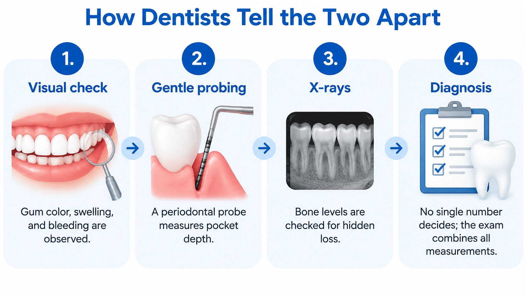
No single number decides the diagnosis. A periodontal examination combines the appearance of the gums, measurements around the teeth, and imaging of the supporting bone. The clinician also considers medical history and other possible explanations for the findings.
The examination in order
- Visual check. The clinician looks at gum color, swelling, and visible bleeding. These findings identify inflammation, but they don't establish support loss.
- Gentle probing. A thin periodontal probe measures the space between the gum edge and tooth at multiple sites around each tooth. Bleeding during probing is recorded alongside pocket depth.
- Attachment measurements. Clinical attachment loss is measured from the cementoenamel junction, the landmark where the crown meets the root, to the base of the pocket. This helps distinguish a swollen gum margin from lost tooth support.
- Support checks. The examination includes recession, tooth mobility, and furcation involvement, meaning damage where the roots of a multirooted tooth divide.
- Imaging. X-rays show bone levels beside the teeth. i-CAT 3D cone beam imaging may be considered when a more detailed view is clinically needed, rather than automatically used for every gum evaluation.
Measurements that separate gingivitis from periodontitis
| Measure | Reading | What It Indicates |
|---|---|---|
| Probing depth | Under 3 mm | Generally normal, according to the Merck Manual's periodontal guidance |
| Probing depth | Over 4 mm | Indicates periodontitis in that guidance, but isn't diagnosed from one reading alone |
| Clinical attachment loss | 1-2 mm | Stage I range, with less than 15% bone loss, under the European Federation of Periodontology classification framework |
| Clinical attachment loss | 3-4 mm | Stage II range, with 15-33% bone loss under that framework |
| Clinical attachment loss | 5 mm or more | Supports a more advanced classification under that framework |
These are clinical benchmarks, not a self-scoring test. Staging also considers the extent of disease, tooth loss related to periodontitis, and treatment complexity.
Why pocket depth needs context
Swollen gums can create a pseudopocket, a deep-looking space without attachment loss. Conversely, recession can expose the root and produce substantial attachment loss even when the remaining pocket isn't especially deep.
For example, a swollen gum margin and a receded gum margin may yield similar probing depths while representing very different levels of support. The fixed crown-to-root landmark helps the clinician interpret that difference.
Disease can also affect one area more than another. Measuring multiple sites prevents an apparently healthy front tooth from standing in for the whole mouth. A plain-language explanation of pocketing in teeth can help patients understand the chart discussed during the appointment.
Causes, Risk Factors and How Common Each Is
What makes one person's gum inflammation remain limited while another develops damage to the tissues supporting the teeth? Bacterial plaque is the shared trigger. This sticky film forms along the gumline and can harden into tartar if it remains. Brushing and cleaning between teeth disrupt plaque; hardened deposits require professional removal.
The amount of plaque alone does not determine the outcome. Tobacco exposure, health conditions, tooth anatomy, home-care habits and family history can all affect risk.
A practical risk checklist
- Smoking: Tobacco exposure raises the risk of periodontal disease. Quitting can reduce this modifiable risk, and professional support may help.
- Diabetes or metabolic disease: These conditions can affect inflammation and disease control. Coordinated medical and dental care can help address risks that can be managed.
- Plaque-retentive anatomy: Crowded teeth, root contours and hard-to-reach spaces can make cleaning less effective. Adapted tools and technique can help reach these areas.
- Inconsistent home care: Plaque left on repeatedly missed surfaces can persist. A steady routine is more useful than brushing harder.
- Family history: Periodontal disease in close relatives signals that closer monitoring may be appropriate. Share that history with your dental professional so it can inform follow-up.
What the population figures mean
Using examination data from 2009-2014, later CDC reporting estimated periodontitis in about 42% of U.S. adults aged 30 and older and about 60% of adults aged 65 and older, as summarized in the CDC's gum disease facts. These are historical population estimates, not predictions for an individual.
An earlier CDC surveillance analysis, based on 2009-2010 examination data, estimated that 47.2% of U.S. adults aged 30 and older, approximately 65 million people, had periodontitis. That estimate excluded gingivitis. Different survey periods, definitions and reporting methods mean the figures are not a simple before-and-after comparison.
Gingivitis and periodontitis are different categories, so a population estimate for one cannot be substituted for the other. Bleeding gums alone do not establish which condition is present. Population figures describe how common disease is; an individual diagnosis depends on clinical examination and measurements.
Can Gingivitis Turn Into Periodontitis
A common assumption is that untreated gingivitis always becomes periodontitis, and that stopping the bleeding means the problem has been cured. Neither conclusion is reliable. Untreated gingivitis can progress, but progression isn't inevitable or identical for every patient.
Tobacco exposure, diabetes or metabolic disease, difficult-to-clean anatomy, the individual inflammatory response, and possible genetic susceptibility all influence the outcome. Successful gingivitis treatment also doesn't provide permanent protection if plaque control and risk management later deteriorate.
A possible pathway, not a fixed timetable
- Healthy gums: Tooth-supporting tissues remain intact, without active gingival inflammation.
- Gingivitis: The gums become inflamed, but effective treatment can return them to health.
- Persistent inflammation: Ongoing plaque and individual susceptibility may allow destructive changes to develop.
- Periodontitis: Attachment and bone loss establish a diagnosis that requires continued risk monitoring.
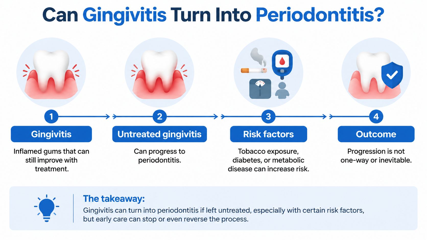
The arrows represent a possible disease pathway, not a deadline or an unavoidable sequence. A discussion of the levels of gingivitis can help explain changes in inflammation, but the transition to periodontitis requires evidence of lost support.
Does the end of bleeding mean a cure?
The answer changes with the diagnosis. When gingivitis resolves and the supporting structures remain intact, the gums may return to health. When periodontitis has been diagnosed, reduced bleeding is encouraging, but it doesn't remove the need for periodontal maintenance.
Consensus literature discussing periodontal risk supports treating a history of periodontitis as a lifelong risk state. A stable patient still needs individualized follow-up because recurrence or further progression remains possible.
Less bleeding is a useful treatment response, not proof that every affected site is stable.
Consider an adult who feels much better after a cleaning and cancels further assessment. If attachment and bone levels haven't been evaluated, the improvement doesn't settle whether the original problem was gingivitis or periodontitis.
A more useful follow-up question is whether the measured sites have stabilized and what maintenance is needed. That keeps the focus on preserving support rather than judging success only by how the gums feel.
Treatment Options for Each Stage
For gingivitis, professional cleaning and an improved daily routine are usually the starting point. Treatment should match the diagnosis, rather than assigning the same procedure to every person with bleeding gums. Home care should address the specific surfaces and spaces where plaque remains.
Treatment options by diagnosis
| Situation | Typical Approach | Goal |
|---|---|---|
| Gingivitis | Professional cleaning and an improved daily routine | Remove plaque and calm inflammation |
| Early periodontitis, stage I to II | Non-surgical scaling and root planing | Clean below the gumline and reduce infection |
| Periodontitis with persistent deep pockets | LANAP laser procedure or osseous surgery, when clinically indicated | Reduce pocket depth; support tissue regeneration where appropriate |
| Gum recession or exposed roots | Soft tissue (gum) grafting | Cover exposed roots and improve comfort where feasible |
| Bone loss after tooth loss | Bone grafting or ridge preservation | Rebuild or preserve bone so implants can be considered |
| Ongoing risk after treatment | Periodontal maintenance visits | Monitor the gums and limit recurrence |
The table is a guide, not a treatment prescription. A periodontist reviews pocket measurements, attachment levels, imaging, medical history, and response to initial care before recommending the next step.
When more involved care may help
Many patients begin with scaling and root planing, which removes deposits below the gumline. Reassessment then helps identify sites where inflammation or deep pockets remain despite initial therapy and improved home care.
Periodontal Associates of Delaware offers non-surgical therapy, LANAP laser gum surgery using the FDA-cleared PerioLase MVP-7, and traditional osseous surgery when clinically indicated. Its services also include soft tissue grafting and bone grafting; when implants are planned after bone loss, X-Nav dynamic surgical navigation may guide placement.
LANAP means Laser-Assisted New Attachment Procedure. It may be appropriate for selected periodontal findings, while osseous surgery may provide better access or address particular bone contours. Neither is an automatic choice based only on a diagnosis label.
Patients considering whether laser gum surgery is worth it benefit from a discussion of candidacy, alternatives, expected recovery, and maintenance rather than a promise of a cure.
For patients anxious about surgery: An evaluation is an opportunity to understand the options, not an automatic commitment to an operation.
Surgery should be discussed in advance, with an explanation of why it's being considered and which comfort options are available. Individual results vary, and a periodontist should assess the approach appropriate to each patient's condition and goals.
Book a Periodontal Evaluation If Warning Signs Appear
Could bleeding gums be gingivitis, periodontitis, or another sign that needs attention? Bleeding alone cannot determine the diagnosis. A dentist or periodontist also checks gum measurements, attachment levels, and bone findings.
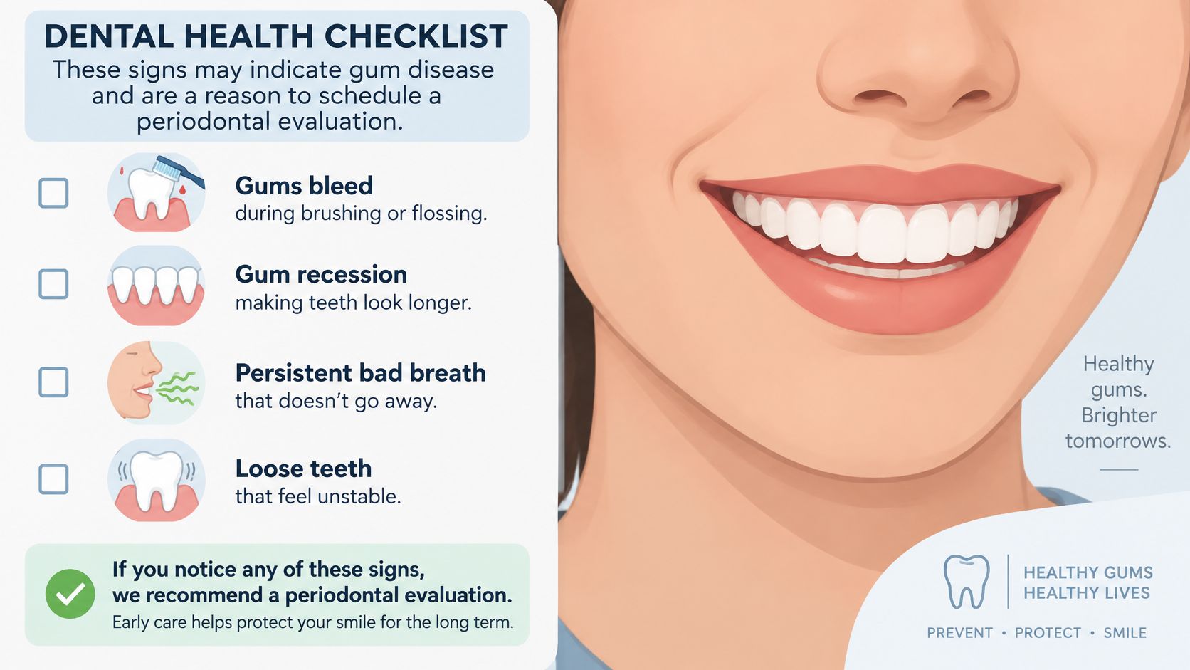
Other changes can help prompt an evaluation: gums receding or teeth appearing longer, persistent bad breath, a loose tooth, or a change in how the teeth meet. Periodontitis may progress with little discomfort, so pain is not a reliable guide to whether tooth-supporting tissues are affected.
When a specialist visit makes sense
Consider seeing a periodontist if a general dentist identifies signs of periodontitis, gum problems persist after professional cleaning, or you want to understand a recommendation for further treatment. A specialist can explain the findings and discuss appropriate options.
Periodontal Associates of Delaware is a board-certified periodontal practice with four Delaware locations: Wilmington, Newark, Dover, and Rehoboth Beach. Its periodontists hold diplomate status with the American Board of Periodontology. A referral is not needed to schedule an appointment.
The practice serves Wilmington, Newark, Dover, Rehoboth Beach, Pike Creek, and Hockessin. Bring any available dental records and a list of medications to help inform the consultation.
Questions to bring to the visit
- Measurements: What pocket depths, attachment levels, and bone findings were recorded?
- Diagnosis: Was periodontitis identified, and what stage was assigned?
- Options: Which treatments fit the findings, and what alternatives are reasonable?
Ask about any concerns before deciding on treatment. Understanding the measurements can clarify whether the issue is gum inflammation or damage to the tissues that support the teeth.
Reversing gingivitis does not erase the possibility of periodontitis later. Keep up with recommended dental care, and ask a periodontist how your findings affect your future risk.
Periodontal Associates of Delaware provides specialist evaluation and individualized care for gum disease. If bleeding, recession, or concerns about bone loss prompt questions, you can request an appointment at one of its four Delaware locations.