A patient often sees the scan body only for a few minutes, after the implant has already healed and the appointment has shifted from surgery to precision. It looks small, but that little part decides whether the lab receives a clear digital map of the implant or a fuzzy guess. For people in Wilmington, Newark, Dover, and nearby Delaware communities who are already uneasy about implant treatment, understanding this step can make the whole process feel more predictable.
Table of Contents
- What a Dental Implant Scan Body Is and Why It Matters
- Component Categories and Materials
- Chairside Workflow From Seating to Scanning
- From Scan Body to CAD/CAM Prosthetic Design
- Accuracy for Single Teeth, Short Spans, and Full Arches
- Variables That Change Scan-Body Accuracy
- How Scan Bodies Fit With CBCT and Surgical Navigation
- Choosing the Right Scan Strategy for the Case
- What to Ask Your Periodontist About Scan-Body Workflows
- Key Takeaways and Next Steps
What a Dental Implant Scan Body Is and Why It Matters
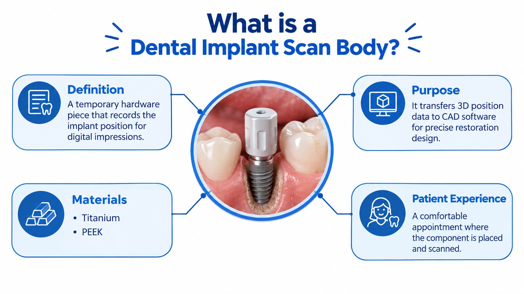
A dental implant scan body is a precision component attached to an implant or implant abutment so a digital scanner can record the implant's three-dimensional position, platform identity, rotational orientation, and emergence profile. In plain language, it gives the computer the exact map of where the implant sits and how it is angled. Without that information, a crown, bridge, or full-arch restoration may look correct on screen and still fit poorly in the mouth.
Why the lab needs more than a picture
The scan body is the handoff point between what is inside the mouth and what is built in CAD/CAM software. That handoff works only when the record is accurate. A systematic review evidence shows that material, geometry, surface characteristics, height, diameter, position, inclination, tightening torque, scanner type, scan strategy, implant angulation, and inter-implant distance can all affect the digital impression (systematic review evidence).
Practical rule: a scan body helps the software read the implant, but it cannot correct poor seating, a rushed scan, or the wrong implant library.
This makes the scan body part of a controlled digital protocol, not a self-contained part that works the same way in every case. The design has to match the implant system, the component has to seat fully, and the scan has to capture it from angles the software can interpret. That is why the seemingly small step carries so much weight. The laboratory is not guessing at implant geometry, it is building from verified geometry when the workflow is done well.
Component Categories and Materials
The first choice is usually material. The two most common options are titanium and polyetheretherketone, or PEEK, and they do not behave the same way inside the scanner. A material comparison study found lower root-mean-square error for titanium scan bodies than for PEEK scan bodies, with p < 0.001, which is one reason clinicians pay close attention to the exact component they select.
Geometry matters as much as the material
Shape changes how the scanner recognizes the component. As that same review noted, short cylindrical forms, cylindrical designs with appropriate dimensions, rigid extensions, simplified reference geometry, and well-defined facets can help the scanner read the scan body more reliably. Some designs are taller, some shorter, and some use asymmetry so the software can orient the implant with less confusion.
That matters because the scan body has to do two jobs at once. It must be easy for the scanner to read, and it must encode the implant position in a way the CAD library can understand. If the geometry is too sparse, too low, or poorly matched to the scan path, the record can become less dependable.
A few practical distinctions help patients understand why one case may use one design and another case a different one:
- Titanium versus PEEK: titanium may offer better reading performance in some laboratory settings, while PEEK can still be appropriate when the system and workflow call for it.
- Short versus tall profiles: shorter bodies can fit better in limited space, but they may be harder to capture when soft tissue covers part of the component.
- Symmetric versus asymmetric designs: asymmetric faces and facets can help the scanner recognize rotation more clearly.
- Single-use versus reusable: reusable components still need checks for wear, cleanliness, and fit, since accuracy depends on what seats in the mouth, not just what the package says.
A scan body also has to match the exact implant-system library. The digital file is not generic. If the library and the physical component do not correspond, the software can build a design that looks plausible but will not seat as intended. That is why these parts are chosen so carefully rather than treated as interchangeable.
Chairside Workflow From Seating to Scanning
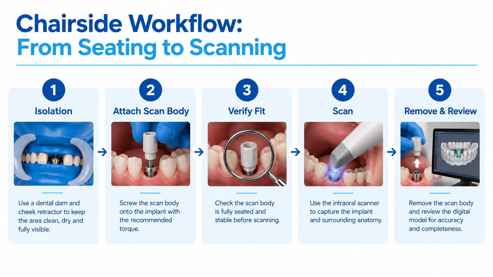
A good scan starts before the scanner ever enters the field. The team isolates the area, manages soft tissue, and seats the scan body on the correct implant-level interface or abutment. The component has to sit fully down and be tightened according to the manufacturer's protocol, because even a small gap can carry into the final design, as noted in clinical guidance.
The chairside sequence is simple, but each step matters.
What the patient may notice in the chair
The scan itself is quiet. A handheld scanner passes over the area while the clinician follows a controlled path, so the software can assemble the images without gaps. Dry, open access helps the process, since moisture, blood, or tissue overgrowth can weaken the read and force the scanner to work harder.
If the connection is hard to see, verification becomes part of the safety check, not an optional extra.
Visual review or radiographic confirmation can help when the interface sits below the gumline or the seating line is difficult to inspect. The aim is straightforward, confirm that the component is fully seated before the record is captured. Once the scan body is recorded, the team can review the model right away and rescan any area that looks incomplete.
The scan body is a precision reference, not a stand-alone gadget. Its value depends on the full digital protocol around it, including how it is seated, how the scan is carried out, and whether the implant-system library matches the physical part. When those pieces line up, the scan gives the laboratory a cleaner starting point and reduces avoidable guessing later.
From Scan Body to CAD/CAM Prosthetic Design
Once the scan is captured, the digital record moves into design software, where the scan body becomes a virtual reference point. The technician sees the implant library, the scanned geometry, and the proposed prosthetic shape all in one file. That library is what lets the software translate the visible component into platform geometry, screw-channel direction, and emergence profile.
A merged workflow can also connect the scan with 3D imaging when implant placement and restoration design are being planned together. In that setting, the scan body helps anchor the restorative plan to the actual implant position, while the 3D image helps show where that implant sits relative to bone and nearby anatomy. The result is a single digital language for surgery, temporization, and the final restoration.
The match has to be exact. A scan body and a library file from the wrong system can create a design that looks convincing on screen but does not correspond to the implant connection. That is why clinicians treat library verification as part of quality control, not a clerical detail.
For patients, the benefit is practical. A cleaner digital handoff can shorten back-and-forth with the laboratory and reduce the risk of avoidable adjustments later. It also means the restoration is designed from recorded geometry, not from a visual estimate made after the fact.
Accuracy for Single Teeth, Short Spans, and Full Arches
A scan body performs best when the case is small and the geometry is easier to capture. For single implants and short-span restorations, scan-body workflows can be comparable to conventional impressions in the right setting, as noted earlier in the article summary of the evidence. A 2023 study found small implant-scan measurement differences around the control planes, with values clustering close to zero and widening as more implants were included, which supports practical accuracy for single implants while showing why more complex cases need more caution (2023 study).
For a single implant, the scan body usually gives the software one clear landmark to read. With a short span, the same workflow can still work well, but each added implant gives the scanner another chance for small orientation errors to build.
A quick comparison of where the workflow tends to help most
| Clinical situation | Typical scan-body outcome | Why caution grows |
|---|---|---|
| Single implant | Often useful and predictable | Less risk of compounded error |
| Short-span bridge | Frequently workable with careful protocol | Small errors can begin to add up |
| Full arch | More challenging and less forgiving | Long spans amplify stitching and orientation problems |
For full-arch and complete-edentulous cases, the margin for error narrows. As noted earlier, accuracy becomes harder to maintain as the scan span and implant count increase, and some laboratory models still favor conventional open-tray techniques over digital methods. One 2020 in-vitro study of complete-arch implant rehabilitation found that digitally redesigned scan-body groups performed better than the conventional designs tested, yet splinted open-tray impressions remained the most accurate for that full-arch model (full-arch review evidence).
The practical lesson is straightforward. Digital accuracy is real, but it depends on the whole protocol, not the scan body alone. Case size, scan path, verification, and implant-system matching all matter more as the span gets longer and the restoration becomes less forgiving.
Variables That Change Scan-Body Accuracy
A scan body is only as accurate as the full protocol around it. The component matters, but so does height, diameter, reference-surface geometry, material, color, surface finish, implant angulation and depth, scanner model, scanning strategy, inter-implant distance, and scan span. A clinical review has also linked these factors to the reliability of the digital record (clinical review). In practice, a fully placed tissue-level implant with little clearance is harder to capture than one that sits more openly and gives the scanner space to read the geometry.
What clinicians watch before scanning
A scan body can only work when the surrounding conditions let the scanner see it clearly.
- Height and exposure: if the body sits too low under tissue, the scanner has less to read.
- Diameter and shape: the component needs enough landmark geometry for software recognition.
- Soft tissue and saliva: blood, moisture, and cheek pressure can blur the record.
- Angulation and spacing: steep implant angles and tight spacing reduce tolerance for small errors.
- Scan path: a scattered scan route raises the chance of stitching errors.
The cleanest way to understand this is simple. The scan body is the reference, and the mouth is the setting around it. If tissue, saliva, or crowding hides part of the component, the digital record becomes less dependable. Careful seating checks, a dry field, and a deliberate scan sequence matter as much as the material or brand.
Clinical takeaway: protocol beats assumption. A good scan body still needs correct placement, clear visibility, and a scanner path that captures enough surrounding landmarks.
Bone loss and soft-tissue changes can make the record harder to read because the neighboring contours are less predictable. In cases where the ridge needs rebuilding before the final prosthetic record is taken, a clinician may also review periodontal bone grafts to understand how the supporting anatomy affects the scan environment.
How Scan Bodies Fit With CBCT and Surgical Navigation
A scan body becomes even more useful when it sits inside a broader digital plan. The surface scan gives the prosthetic reference, and i-CAT 3D cone beam imaging shows the bone, sinus, and nerve anatomy that shape implant placement. When those datasets are merged, the team can plan a position that works for both the restoration and the underlying anatomy.
That coordination matters clinically because implant placement and prosthetic design are linked. If the implant ends up where the digital plan assumed it would be, the final restoration is more likely to follow the intended screw channel and emergence profile. If the surgical position drifts, the prosthetic plan may need to be adjusted later.
X-Nav dynamic surgical navigation can use the merged records to guide implant placement in real time. That does not make the scan body “accurate” by itself, but it helps keep the surgery aligned with the restorative plan that was built from the scan data. In a patient's chart, that often means fewer surprises during placement and a cleaner path toward temporization.
For cases that involve limited vertical bone, sinus proximity, or planned augmentation, the imaging and scan-body record need to stay coordinated from the start. That is why many planning conversations include the digital record and, when needed, reference material on sinus lift procedures performed at the same time as implant placement. The goal is consistency, not complexity for its own sake.
Choosing the Right Scan Strategy for the Case
A scan body works best when the whole digital path fits the case. For a single tooth or a short span, the implant is usually easier to expose, the scan is easier to capture, and the implant library can be matched with more confidence. As the span grows, access gets tighter, angulation matters more, and the team may need a conventional or hybrid workflow.
That choice is shaped by anatomy, visibility, scanner performance, and the restorative goal. It also depends on whether there is enough room for the restorative components to be captured cleanly, which is why dental implant space requirements are part of the planning conversation.
The evidence base is still mostly laboratory-focused. As the review covered earlier in this article found, 16 eligible studies examined factors influencing intraoral implant scanning, and a 2023 review included 28 in-vitro studies from 2019 through 2023, with the strongest results in single implants and short spans (review evidence). That supports digital care in the right situations, but it does not make every mouth a digital case.
A practical way to sort the options is straightforward:
- Digital scan-body workflows fit best when the implant is easy to access, the scan span is short, and the library match is confirmed.
- Conventional impressions still matter when the span is long, the implants are difficult to visualize, or stitching is less dependable.
- Hybrid workflows help when the team wants the advantages of digital capture without forcing every step into one method.
Patients can ask direct questions at the consultation, which method fits the case, why that method was chosen, and how the scan body, scanner, and library will be checked before fabrication. That judgment depends on anatomy, access, scanner performance, and the restorative goal rather than a marketing label.
What to Ask Your Periodontist About Scan-Body Workflows
A good consultation should leave the patient with a clear plan, not just a technical label. The most useful questions are straightforward: which impression method fits the case, which scan-body system matches the implant, how seating will be verified, and how the digital plan connects with imaging and navigation. Those questions help the patient see whether the workflow is being matched to the anatomy or forced onto it.
A short checklist for the visit
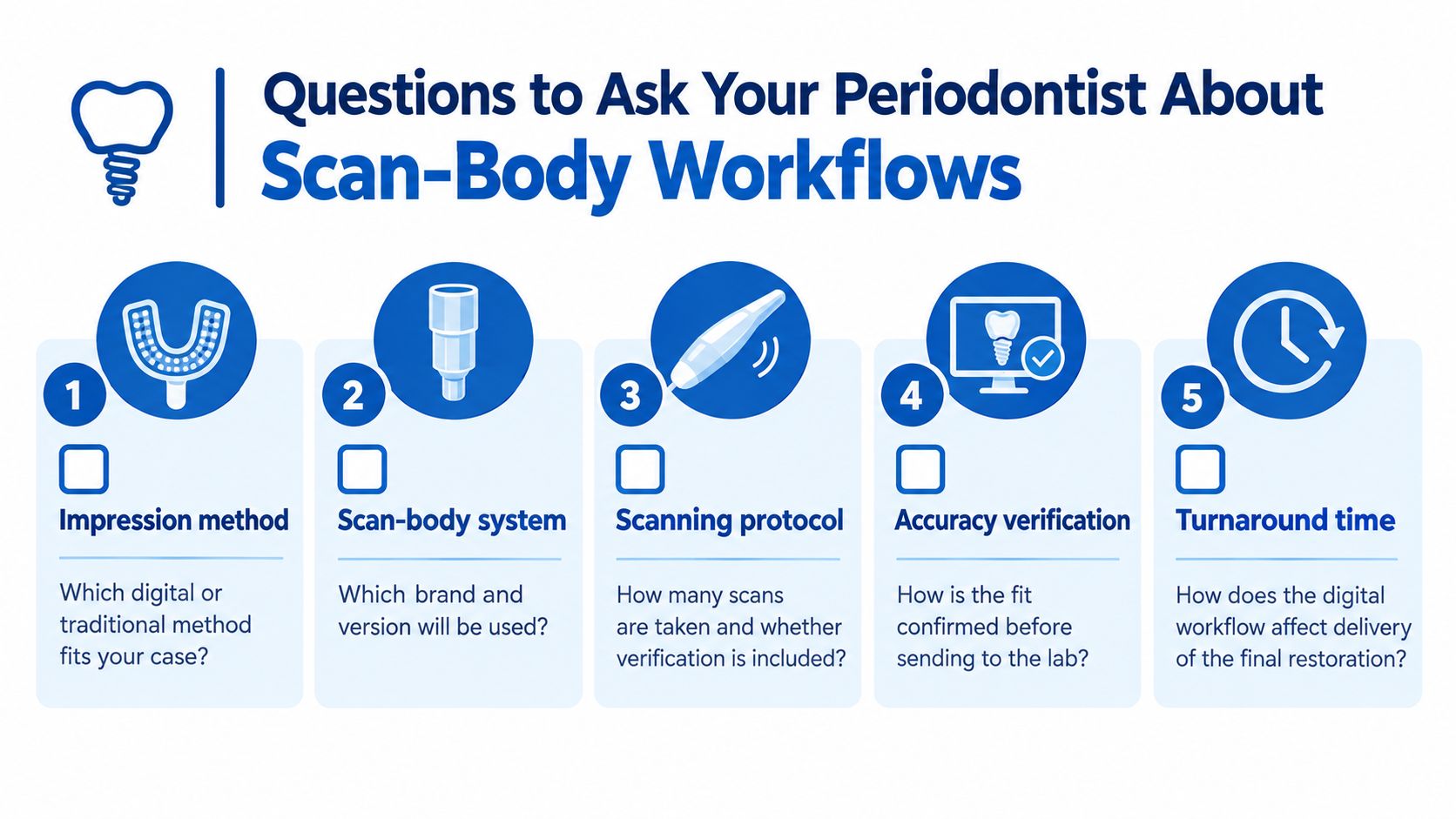
- Impression method: Which digital or traditional approach is best for this mouth?
- Scan-body system: Which brand and version will be used, and why?
- Scanning protocol: How is seating checked, and how many scans are taken?
- Accuracy verification: How is fit confirmed before the file goes to the lab?
- Turnaround time: How does the workflow affect the final restoration schedule?
Patients in Wilmington, Newark, and Dover can ask for the office that is closest to them, since care is available across all three Delaware locations. Individual results vary, and no outcome is guaranteed, so a personalized evaluation from a periodontist remains the right starting point before any implant recommendation is made.
Key Takeaways and Next Steps
A dental implant scan body is a precision interface, not a generic marker. Its job is to transfer implant position, rotation, and platform details into the digital record that guides the restoration.
Accuracy depends on the whole protocol, especially design, seating, scan strategy, and implant-system library matching. The evidence is strongest for single implants and short spans, while full-arch cases remain more demanding.
Patients across Wilmington, Newark, Dover, and surrounding Delaware communities can use this knowledge to ask better questions and choose a workflow that fits their anatomy, comfort level, and long-term restorative goals.
Periodontal Associates of Delaware provides periodontal evaluation, implant planning, and advanced digital tools that may support a precise restorative workflow, including scan-body records, 3D imaging, and surgical navigation. To learn whether this approach fits your situation, visit Periodontal Associates of Delaware and request an appointment at the Wilmington, Newark, or Dover office that's most convenient for you.