
You catch a gray, brown, blue, or black rim on the gum of one tooth while the rest of the smile looks unchanged. It may sit beside a filling, a crown, or a tooth that has started to bleed when you floss. Dark gums around a tooth raise a fair question, and the first fear is usually gum disease.
That fear is understandable, and it is also incomplete. A patch around a single tooth is a different problem from gums that have always been evenly brown. Color can point toward a metal stain, natural pigment, inflammation, or a lesion that needs a tissue sample. Color cannot prove which one it is.
The pages ahead separate those patterns, show how each one forms, and spell out when to measure, image, or sample tissue before any cosmetic plan. A periodontist confirms the match. Individual results vary, and a written description can't diagnose the spot.
Table of Contents
- Dark Spot Patterns and Likely Causes
- How Each Cause Forms and Looks
- What a Periodontal Exam Measures
- Changes That Should Not Wait
- Treatment Options Matched to the Cause
- Book a Visit at the Nearest Office
Dark Spot Patterns and Likely Causes
A dark area around one tooth isn't automatically periodontal disease. A clinical review of oral pigmentation places physiologic melanin pigmentation, an amalgam or endodontic-material tattoo, medication-induced melanosis, systemic disease, melanocytic lesions, and oral melanoma on the same list. Exogenous dental pigments can migrate through bone or soft tissue near the tooth apex and often leave a localized gray-blue or black mark.
Match one row before any whitening idea. A periodontist should confirm the match, and individual results vary.
Localized dark gum patterns and next steps
| Look | Likely cause | Often found with | Next step |
|---|---|---|---|
| Even brown gum, stable for years | Physiologic melanin | Flat surface, often more than one area | Document and monitor |
| Sharp blue-gray or black patch by a filling or crown | Amalgam or endodontic-material tattoo | Prior restoration or root canal | Periapical x-ray; biopsy if unclear |
| Brown attached gum, often lower front teeth | Smoker's melanosis | Current tobacco use | Full exam; do not assume tobacco |
| Dusky, purple, or black swollen margin | Inflammation or tissue breakdown | Bleeding, pus, pain, or a deep pocket | Periodontal measurements and care |
| Broader darkening after a medicine | Medication-related melanosis | Drug history | Clinician review and rule-out of other causes |
| Solitary uneven spot that is new or changing | Melanocytic lesion or oral melanoma | Irregular border, ulcer, or loose tooth | Prompt biopsy |
Physiologic melanin is usually flat, even, and unchanged for years on attached gum. It often appears in more than one area. That pattern is a pigment map, not a failing tooth.
An amalgam or endodontic-material tattoo is a sharp gray-blue or black macule beside a restored tooth. Particles can move through nearby soft tissue or bone. When the source is unclear, a periapical radiograph can help identify metal-related pigmentation or a restorative-material source. If the film and the history still don't line up, the next step is a biopsy, not a bleach tray.
Medication-related melanosis follows a drug history and is often wider than one tooth. Smoker's melanosis favors attached gum, especially the lower front teeth. A new solitary spot shouldn't be blamed on tobacco by default.
Inflamed or breaking-down tissue looks dusky, purple, or black and comes with swelling, bleeding, pus, pain, or a deep pocket. That pattern needs measurements. Early bleeding and puffiness sit among the levels of gingivitis, and a photo still can't finish the chart.
A melanocytic lesion or oral melanoma is solitary, uneven, changing, raised, or ulcerated. Observation fits only when the source is clear and stable. An unexplained or changing lesion generally needs tissue biopsy to exclude malignancy rather than cosmetic treatment alone.
Practical rule: Note when the rim appeared, whether it grew, and which tooth has a filling or root canal. Those three facts steer the visit.
The same visit should also record symmetry, borders, surface changes, tobacco and medication exposure, and whether the spot is tied to inflammation, bleeding, pain, or ulceration.
How Each Cause Forms and Looks
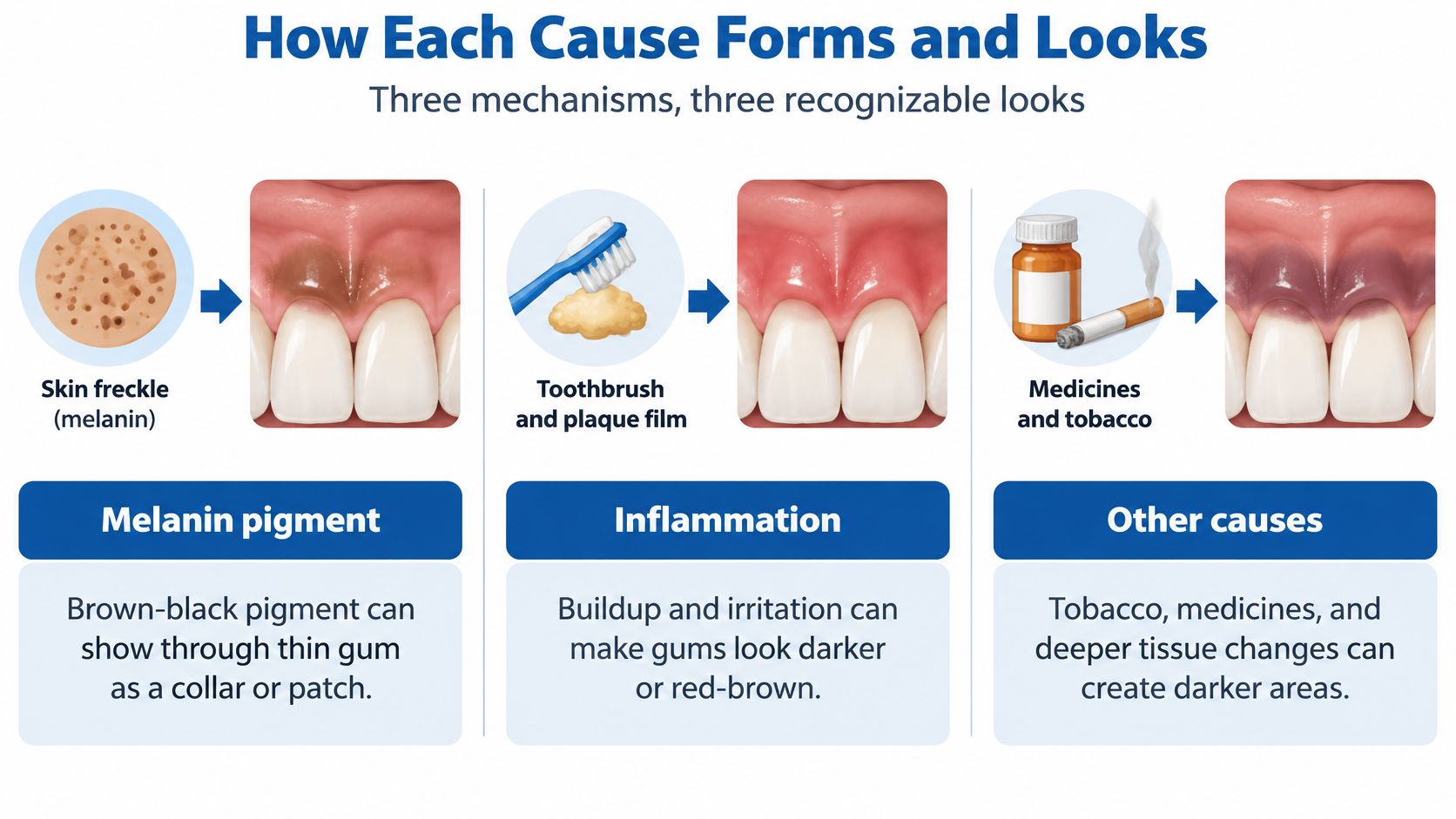
Three processes can leave dark gums around a tooth, and each one has a look you can recognize.
Melanin that shows through thin gum
Melanin is the same brown-black pigment found in skin freckles. Thin gum can show it as a collar or a patch. Many adults carry some natural darkness without disease. The surface stays flat, and the shade doesn't arrive overnight beside one back tooth.
Tobacco can push that pigment higher. In one study, visible gingival melanin pigmentation occurred in 27.5% of current smokers compared with 8.6% of people who had never smoked, and the difference was statistically significant, as reported in a 2024 review of gingival melanin pigmentation. Nicotine and benzopyrene may stimulate melanin production. The color often sits on attached gum, particularly around the lower front teeth.
Say a current smoker has brown attached gum on the lower front teeth, no raised edge, and no ulcer. That story fits stimulated melanin. It doesn't explain a brand-new patch beside one molar.
Metal that stays like a permanent bruise
An amalgam tattoo is a localized deposit of dental-metal particles in the gum or nearby oral tissue. A clinical overview of amalgam tattoo explains that these particles may become embedded during a filling or other dental procedure and can create a persistent blue, gray, or black patch. Examination comes first. Dental radiographs may show radiopaque metal particles. If imaging doesn't show particles, or if the history is unclear, biopsy may be recommended, because an isolated pigmented spot can resemble a melanocytic lesion or oral melanoma.
Picture a painless blue-gray patch beside a silver filling. It hasn't ulcerated, and it hasn't spread across the arch. Fine metal from a filling, a crown edge, or root-canal material lodged in the tissue and stayed. That isn't a bruise that fades in a week.
Blood and fluid, not extra pigment
Inflammation is a blood and fluid problem. Swollen gum holds more blood, so the margin may look purple. Tissue that is breaking down may look gray-black. Pain, bleeding, a bad taste, food trapping, or a loose tooth usually travels with that change.
A dark swollen margin that bleeds when you floss, traps food on one side, or sits on a tooth that rocks is a disease story, not a freckle story.
A common background finding doesn't explain a brand-new patch beside one back tooth. Mixed stories still need an exam.
What a Periodontal Exam Measures
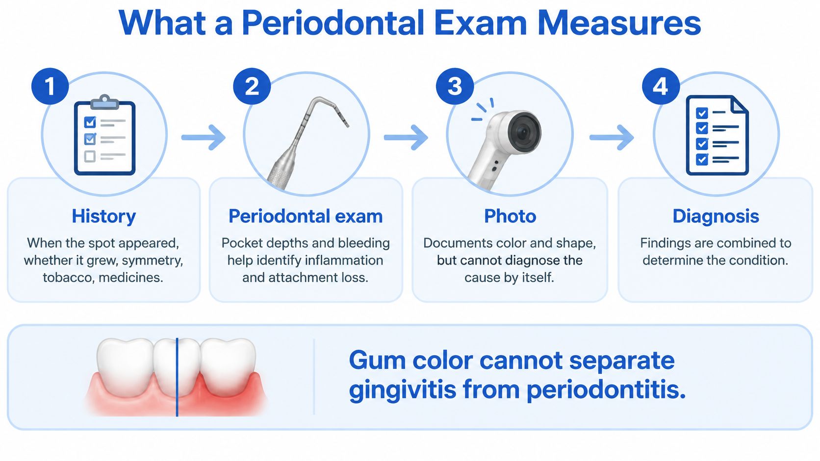
Gum color can't separate gingivitis from periodontitis, so the visit records more than a photo. A periodontal disease reference from the National Library of Medicine states that gingival appearance alone cannot make that split. Gingivitis is inflammation without attachment loss. Periodontitis includes attachment loss and may show alveolar bone loss on films.
The visit in order
- History comes first. The clinician asks when the spot appeared, whether it grew, and whether the other side matches. Tobacco, medicines, old fillings, and root canals go on the chart, along with bleeding, pain, or an ulcer.
- The look is described, not diagnosed. Borders, mixed colors, and a flat or raised surface are written down. The note also says whether the mark is tied to one tooth.
- Six sites around that tooth get measured. Plaque and calculus, bleeding on probing, pocket depth, clinical attachment level, recession, and mobility are checked. Those numbers, not the shade, show whether the gum has only swollen or has lost attachment. Deep readings are the same problem described in pocketing in teeth.
- Periapical or bitewing images come next. A bright metal speck can support a tattoo. Bone loss on the film supports periodontitis. Cone-beam imaging such as i-CAT is reserved for selected bone or implant questions, not a routine color screening.
- The decision is monitor or biopsy. A stable, explained cause can be photographed and watched. An unexplained cause gets a tissue sample.
A dark margin with edema, bleeding, pus, deep pockets, recession, or radiographic bone loss may reflect inflammation or disease-related tissue change. A stable, sharply localized blue-gray macule next to a restored tooth may favor an exogenous tattoo.
Asymmetric, enlarging, irregularly bordered, ulcerated, or otherwise unexplained pigment warrants prompt specialist review.
That review can happen at a Wilmington, Newark, or Dover office. The goal of the first visit is a clear record, not a cosmetic plan.
Changes That Should Not Wait
A flat blue-gray spot that has sat beside an old filling for years isn't the same problem as a new dark area that is changing. Waiting for the next cleaning, or booking a whitening visit first, rests on the wrong assumption. Stable and unchanged can be watched. New and changing should not.
Oral melanoma is uncommon, but it belongs on the list. A faculty review of oral melanoma says it must be considered when a dark gum lesion is new, solitary, changing, irregular, raised, bleeding, ulcerated, or associated with tooth loosening. Appearance alone cannot establish the diagnosis. Biopsy is the most reliable confirmatory test when malignancy is suspected.
Warning features in plain words
- Asymmetry: One side of the spot doesn't match the other.
- Irregular borders: The edge is jagged or fades in uneven steps.
- Uneven color: Brown, black, blue, or red sit inside the same patch.
- Size: A diameter greater than 6 millimeters is a reported warning feature.
- Elevation: The surface is raised rather than flat.
Advanced oral melanoma may add spontaneous bleeding, pain, ulceration, or mobility of nearby teeth. A dark swelling that also drains or hurts can overlap with a periodontal gum abscess, which is another reason not to scrub the area or wait it out.
Don't rub the spot with a hard brush, and don't seek color removal first if those features are present. A same-week periodontal visit is the practical action. Most dark spots aren't melanoma. The list is a reason to be seen, not a self-diagnosis. Individual findings vary.
Treatment Options Matched to the Cause
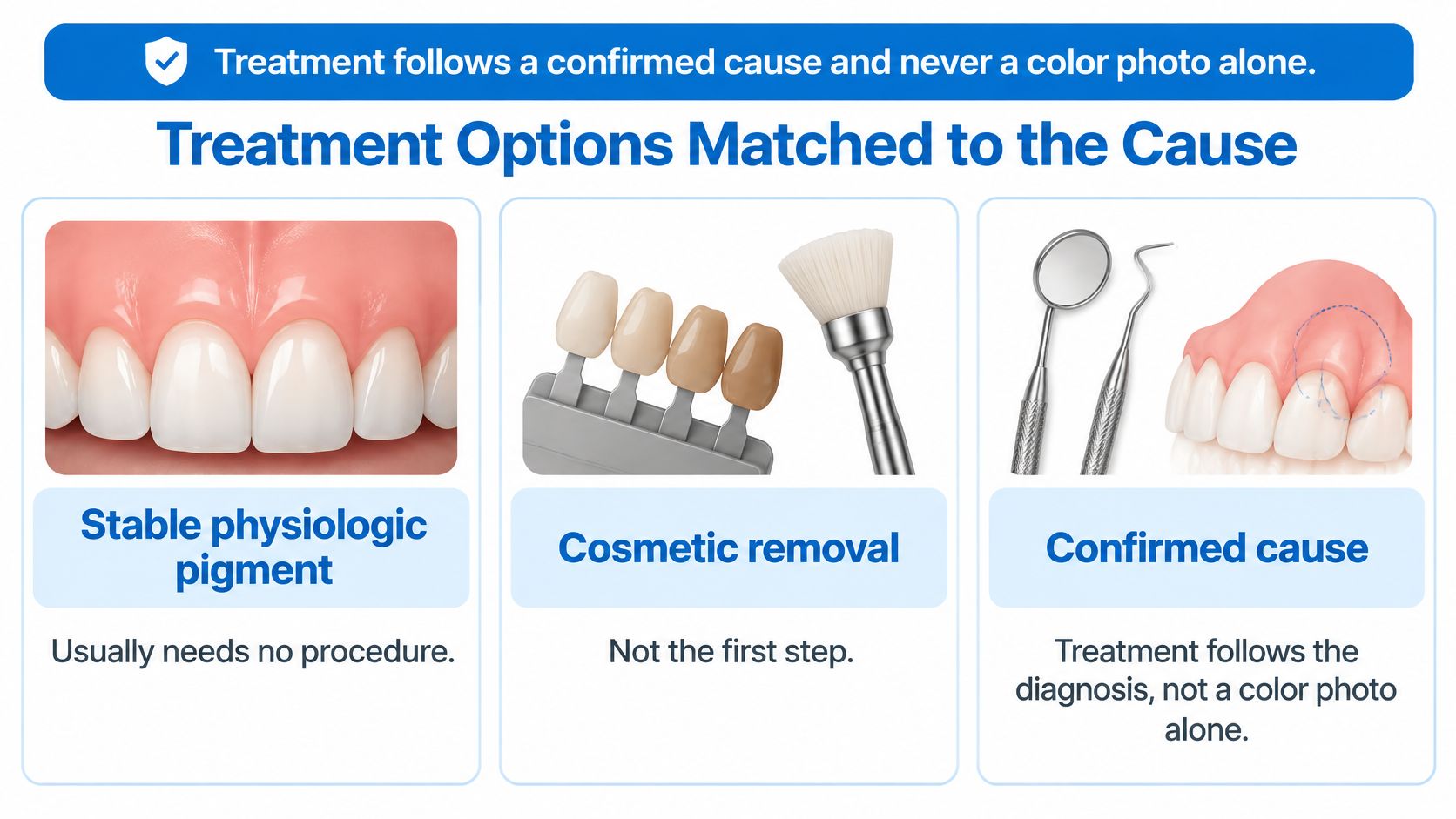
Treatment follows a confirmed cause and never a color photo alone. Removal is a fair question, and it is the wrong first move when the cause is still a guess.
Care after the cause is confirmed
| Confirmed cause | Care that may be offered | Limit to expect |
|---|---|---|
| Physiologic melanin | Observation and photos | Color may remain; no disease treatment needed |
| Amalgam or endodontic-material tattoo | Observation after diagnosis | The patch may persist |
| Smoker's melanosis | Quitting tobacco plus monitoring | Darkening may lessen gradually |
| Gingivitis or periodontitis | Cleaning, maintenance, LANAP or surgery when indicated | Inflammation may improve; results vary |
| Unexplained or changing lesion | Biopsy before other treatment | Looks alone cannot exclude melanoma |
| Tooth that cannot be saved | Site preservation and possible implant planning | Only when the site has enough healthy bone and soft tissue |
Stable physiologic pigment usually needs no procedure. Cosmetic removal isn't the first step. A confirmed amalgam tattoo is usually observed, because the color often stays. A confirmed metal stain isn't usually lightened with whitening products or stain removers. Don't use bleaching gels or abrasive pastes on a dark gum spot before it is evaluated. A cosmetic request is discussed only after the cause is known. The periodontist can explain whether any removal is appropriate, what it may involve, and what tradeoffs exist, including a change in gum shape or healing.
Smoker's melanosis may lighten after quitting. Many patients see only gradual change, not a guaranteed fade.
When the cause is gum disease
For gingivitis or periodontitis, care may include scaling and root planing and periodontal maintenance. When pockets and bone loss fit, LANAP laser gum surgery with the FDA-cleared PerioLase MVP-7 may be discussed as a minimally invasive option. Traditional osseous surgery may be used when it is clinically indicated. Many patients feel anxious about surgery. Comfort and healing vary. No outcome, cure, or cost is promised.
If disease has loosened a tooth that can't be kept, the plan may shift to extraction-site preservation. When an implant is appropriate, i-CAT 3D cone beam imaging and X-Nav dynamic surgical navigation may be used for placement. An unexplained or changing dark spot gets a biopsy before any cosmetic or laser color treatment.
Individual results vary, and a periodontist should match the option to the measurements, not to a phone photo.
Book a Visit at the Nearest Office
Book soon if the dark area around the tooth is new, one-sided, growing, sore, bleeding, ulcerated, or linked to a loose tooth. A long-stable spot still deserves a baseline photo and measurements so a later change is obvious.
Periodontal Associates of Delaware is a board-certified periodontal practice with offices in Wilmington, Newark, and Dover. The team cares for adults across New Castle and Kent Counties, including Pike Creek, Hockessin, Bear, Middletown, Dover, and nearby towns. The visit can sort pigment, metal stain, inflammation, and lesions that need biopsy. It can also plan gum treatment if pockets or recession are present.
Many patients feel anxious about surgery. The exam itself is a conversation and a set of measurements. Any procedure is discussed only if the findings support it. Individual results vary, and this article can't diagnose a spot from a description.
Request an appointment at the nearest of the Wilmington, Newark, or Dover offices through the online appointment page.
Periodontal Associates of Delaware offers board-certified periodontal care for a single dark patch, gum disease, recession, and teeth that may need implants, with offices in Wilmington, Newark, and Dover. A visit can measure the tooth, image the area when it is indicated, and decide whether the spot should be watched or biopsied before any cosmetic step. Request a time at the nearest office through Periodontal Associates of Delaware.