Seeing a photo of gums looking calmer, tighter, or less swollen can feel reassuring, especially when bleeding, bad breath, or loose teeth have been on the mind for months. The hard part is that a good-looking photo does not automatically prove healing, and it definitely does not prove regeneration. Laser gum treatment before and after should be read like a periodontal chart, not like a cosmetic makeover.
A careful reading starts with four questions. Did the pocket get shallower, did the tooth gain real attachment, did recession change, and did bleeding improve? Those are different things, and they do not always move together. A small, honest change on a chart can mean more than a dramatic smile photo.
Table of Contents
- What Before and After Really Means for Laser Gum Treatment
- How Laser Periodontal Therapy Actually Works
- Reading the Numbers Behind Before and After Photos
- Realistic Patient Stories and What to Expect
- Laser Therapy Compared With Other Periodontal Options
- Recovery Timelines and Daily Life After Treatment
- Common Misconceptions Worth Addressing Before You Decide
- Your Next Steps and What to Ask at a Consultation
What Before and After Really Means for Laser Gum Treatment
The most common mistake is treating gum photos like skin photos. Gums can look less red and puffy after treatment because inflammation dropped, but that does not always mean the supporting tissues rebuilt in a lasting way. In periodontal care, appearance is only one clue, and it's often the least reliable one.
What the picture can show, and what it can't
A before photo may show swollen margins, bleeding, and deep shadows around the teeth. An after photo may show a cleaner gumline and less redness. That improvement can reflect debridement, better home care, healing, and maintenance, not just the laser itself.
Practical rule: if a case relies on a single photo, it proves almost nothing. If it includes baseline charting and follow-up measurements, it becomes clinically useful.
Readers also need to separate smaller pocket depth from true rebuilding of support. A pocket can become shallower because swelling goes down and tissue tightens. That is helpful, but it isn't the same as new attachment or bone return.
The rest of this article uses four lenses to judge any before-and-after case the right way, probing depth, clinical attachment level, recession, and bleeding on probing. With those four numbers in mind, a patient can read an image the way a periodontist does, with calm skepticism and clear expectations.
How Laser Periodontal Therapy Actually Works
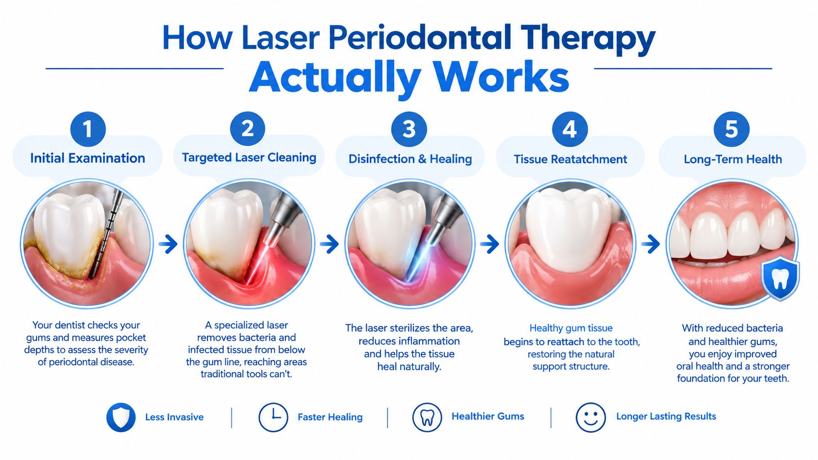
LANAP is not a vague “laser on the gums” concept. It is a defined periodontal protocol using a 1064-nm Nd:YAG laser, delivered through the PerioLase MVP-7, and it's used in a specific sequence rather than as a general heat source. That matters because wavelength, power, and technique change what tissue is affected and what stays intact.
What the laser is doing inside the pocket
Inside an inflamed periodontal pocket, the laser energy is used to target diseased tissue and disrupt the bacterial environment while preserving more of the healthy surrounding tissue. Think of it less like a knife and more like a very controlled pulse of energy that works in a narrow space. The pocket is then cleaned and stabilized so healing can occur.
That process can help shrink inflamed tissue, but it can't be treated as a guarantee of bone regrowth on its own. Bone support, tooth stability, and long-term attachment depend on the full periodontal picture, not one device or one appointment. The diagnostic side matters, which is why 3D cone beam imaging can be part of the workup when the anatomy, bone pattern, or implant planning needs more detail.
For readers who want the technology laid out in one place, Periodontal Associates of Delaware describes its imaging and treatment technology on its technology page. The key point is simple, though. Different lasers are not interchangeable. Diode lasers, CO2 lasers, Er:YAG systems, and Nd:YAG protocols each behave differently, so a meaningful recommendation depends on the exact wavelength and treatment plan.
Why protocol matters more than the word “laser”
Two patients can hear the same phrase, “laser gum treatment,” and receive completely different care. One may have a pocket decontamination protocol, another may have LANAP, and another may need conventional surgery. That's why the machine name alone never tells the full story.
A proper consultation should answer whether the goal is infection control, pocket reduction, attachment improvement, or implant planning. Without that distinction, the before-and-after photo can be persuasive while still being incomplete.
Reading the Numbers Behind Before and After Photos
A credible before-and-after case should show four things, and the sequence matters. First, probing depth tells how deep the pocket is. Second, clinical attachment level shows how much support the tooth has lost or regained. Third, recession shows where the gum margin sits. Fourth, bleeding on probing shows active inflammation.
How to read the measurements like a clinician
In one prospective study of full-mouth LANAP in 8 adults, mean probing depth changed from 4.62 mm to 3.14 mm, while mean clinical attachment level improved from 5.58 mm to 4.66 mm. Mean recession also increased, from 0.86 mm to 1.52 mm. Those numbers matter because they show that a smaller pocket can come from both true improvement and tissue remodeling, not from a simple “regrew the bone” story. Clinical data from the 9-month LANAP study
The most useful habit is to ask for baseline and follow-up at comparable intervals. If someone shows only a photo, that image may be suggestive. If they show charting, recession, and bleeding status, the case becomes far more believable.
Simple checklist: look for pocket depth, attachment level, recession, and bleeding. If one of those is missing, the before-and-after story is incomplete.
| Measurement | What It Tracks | How It Is Taken | What a Meaningful Change Looks Like |
|---|---|---|---|
| Probing depth | How deep the gum pocket is around the tooth | Measured with a periodontal probe at multiple sites | Shallower pockets suggest less inflammation and easier maintenance |
| Clinical attachment level | How much support the tooth has lost or regained | Measured from a fixed reference point on the tooth | An improvement suggests better tooth support, not just calmer gums |
| Recession | Where the gum margin sits on the tooth | Measured from the gumline to the reference point | An increase can happen even when pockets improve |
| Bleeding on probing | Whether the tissue is still inflamed | Noted when gentle probing causes bleeding | Less bleeding usually suggests better control of inflammation |
The table is more trustworthy than a close-up smile photo because it measures change instead of style. That's the standard readers should ask for every time.
Realistic Patient Stories and What to Expect
A patient with deep pockets often starts with a full periodontal exam, charting, and imaging. In some cases, the visit also includes 3D imaging to map bone shape and identify areas that need a more precise plan. The treatment then follows the diagnosis, not the other way around.
What a typical journey looks like
One common path begins with inflamed, bleeding gums and a discussion of whether LANAP, non-surgical therapy, or surgery makes the most sense. Another starts with advanced disease, where the largest numeric change tends to happen because the baseline pockets are deeper. A third begins with a tooth that can't be saved, so the conversation shifts toward extraction and later implant planning.
The first week usually focuses on healing, gentle eating, and avoiding unnecessary irritation. After that, the comparison happens later, when the chart is repeated and the doctor compares the new numbers with the baseline. That follow-up is where the “after” becomes meaningful.
Deep pockets can improve dramatically on paper, but that doesn't mean every site behaves the same way. Smoking, diabetes, plaque control, and maintenance all shape the result.
One Delaware practice that integrates periodontal care with imaging and implant planning is Periodontal Associates of Delaware, which serves Wilmington, Newark, and Dover. A patient with a mix of gum disease, recession, and missing teeth may need a plan that combines periodontal therapy with restorative planning instead of chasing a single dramatic photo.

The most honest before-and-after stories are usually the least flashy. They show a diagnosis, a treatment plan, a healing period, and a measured recheck. That is the kind of progress patients should ask for.
Laser Therapy Compared With Other Periodontal Options
Laser treatment has a place in periodontal care, but it isn't the only option and it isn't automatically the right one. A 2024 review of 266 patients comparing LANAP and SRP found no significant overall difference in the main clinical measures, even though some individual studies favored LANAP for certain pocket-depth outcomes and one study showed attachment gain in its favor. That's an important reminder that both approaches can help, but the outcome depends on the case. 2024 review of LANAP versus SRP
Where each option tends to fit
Scaling and root planing is the standard deep-cleaning approach and may be enough for many cases that aren't too advanced. Traditional flap or osseous surgery may be more appropriate when access to the roots or bone is limited. Laser protocols may be appealing when the treatment goal is less invasive pocket management, but they still need proper case selection.
If teeth can't be saved, the discussion shifts to replacement planning rather than continued pocket reduction. That is where i-CAT 3D imaging and X-Nav dynamic surgical navigation can support implant planning by showing bone volume and guiding placement with more precision.
For readers comparing gum grafting, recession care, and related options, the practice's gum graft alternatives resource can help frame the conversation in practical terms. The right answer usually depends on pocket depth, bone pattern, medical history, and patient preference, not on marketing language.
Recovery Timelines and Daily Life After Treatment
Most patients want the same answer in plain language. Is recovery easier? Often, yes, but it's not pain-free and it's not effortless. Early discomfort can be mild to moderate, and many people need to slow down for the first day or two.
What the first days usually feel like
The early phase often includes soft foods, reduced chewing on the treated side, and a short break from brushing directly on the area. Activity is usually limited for about 48 to 72 hours, then patients gradually return to normal routines as comfort allows. That doesn't mean the tissues are finished healing, only that the most sensitive stage has passed.
A 2024 review of diode-laser-assisted flap surgery found that pain reduction was most evident on the first postoperative day, not consistently later. That lines up with the practical message patients need to hear, laser care may feel easier, but it's still periodontal treatment, not a spa procedure. 2024 review of diode-laser-assisted flap surgery
Maintenance protects the result. Without it, inflammation can return and the after-photo stops telling the same story.
The long-term schedule matters just as much as the treatment day. Periodontal maintenance is usually repeated at regular intervals, often around every three to four months, because the mouth keeps changing after the procedure. The final after-image a patient sees months later reflects both the therapy and the upkeep that followed it.
Common Misconceptions Worth Addressing Before You Decide
The biggest myth is that every laser procedure is gentle and painless. That isn't true. Some patients are comfortable quickly, others still feel soreness, and the experience varies with disease severity, tissue condition, and the exact protocol used.

What the evidence actually supports
Another misconception is that a laser automatically eliminates follow-up care. It doesn't. Periodontal disease is driven by bacteria, plaque, and risk factors over time, so maintenance is part of the treatment, not an optional add-on. The third myth is that all lasers are the same, when in fact wavelength and protocol can change the outcome and the tissue response.
The American Academy of Periodontology's evidence review found that laser evidence is inconsistent, with some promise in certain deep-pocket cases but not a blanket proof that laser therapy is superior across the board. It also warns that inappropriate selection or settings can damage periodontal tissues. AAP evidence review on lasers in periodontics
The safest way to think about before-and-after photos is this. They can be encouraging, but they don't prove regeneration by themselves. They should lead to a conversation about measurements, diagnosis, and maintenance, not to a promise.
Your Next Steps and What to Ask at a Consultation
A patient doesn't need to become a periodontist to make a smart decision. A short checklist is enough. Ask for baseline probing depths, clinical attachment levels, recession, bleeding status, the proposed laser wavelength and protocol, and the maintenance schedule that follows treatment.
Also ask how smoking, diabetes, and bone loss affect the plan. If tooth replacement may be part of the picture, ask whether imaging and implant planning are needed before any gum procedure starts. For patients comparing treatment costs and timing, laser gum treatment cost guidance can help frame the financial side of the decision.
Periodontal Associates of Delaware serves Wilmington, Newark, and Dover, and its board-certified team uses LANAP with the PerioLase MVP-7, i-CAT 3D imaging, and X-Nav dynamic navigation when implant planning is part of care. The most useful before-and-after for a specific mouth comes from a one-on-one evaluation, not from a generic online photo. Individual results vary, and a consult with a periodontist is the right next step.
Periodontal Associates of Delaware helps patients in Wilmington, Newark, and Dover understand what laser gum treatment can and can't do before they commit to care. A consultation can connect the photo, the chart, and the long-term plan so the result is measured clearly. Request an appointment at the nearest office through Periodontal Associates of Delaware, or use the appointment page to schedule a visit that fits your location and concerns.