A patient may notice it while brushing: the gum near a lower front tooth seems to be disappearing, the root looks more exposed, and a tight band of tissue appears to pull whenever the lip moves. That discovery can be unsettling, especially when the area also feels sensitive or difficult to clean.
The band may be a frenum, and the central question is reasonable. Can a small strip of tissue contribute to gum recession, and does cutting it repair the lost gum? A frenectomy may remove one source of tension, but gum recession usually has several possible causes. The right plan may involve hygiene improvement, periodontal therapy, frenectomy, gum grafting, or a combination of treatments.
Patients in Wilmington, Newark, Dover, and surrounding communities can receive periodontal evaluations across all three Delaware offices. Modern care focuses on understanding the cause before recommending surgery. For patients also concerned about nighttime clenching or grinding, the relationship between those habits and gum changes is addressed in this guide to grinding teeth and receding gums.
Table of Contents
- Why Your Gum Line Might Be Pulling Away
- What a Frenum Is and When It Becomes Aberrant
- How a Tight Frenum Contributes to Gum Recession
- When a Frenectomy Is Actually Indicated
- The Frenectomy Procedure at a Periodontal Practice
- What to Expect After a Frenectomy and What It Can Achieve
- Your Next Step Toward Healthier Gums
Why Your Gum Line Might Be Pulling Away
A receding gum line isn't always caused by one dramatic event. A patient may brush carefully, notice no major pain, and still see a narrow area of root exposure near the lower front teeth. When the lower lip is lifted, a taut strip of tissue may become visible between the lip and gum. That strip can feel as though it pulls on the gum every time the mouth moves.
The tissue may be a frenum with an attachment that sits unusually close to the gum edge. In that situation, the frenum can act like a rope tied too near the edge of a tent. Each movement doesn't necessarily cause immediate damage, but repeated tension may make it harder for delicate gum tissue to remain stable, particularly when plaque, inflammation, tooth position, or brushing trauma also plays a role.
A useful expectation: Frenectomy can address a pull on the gum. It can't automatically rebuild gum that has already been lost or correct every other factor affecting the area.
A periodontal evaluation helps separate those possibilities. The clinician examines the frenum, measures the recession, checks the depth of the gum pockets, reviews plaque and inflammation, and evaluates the position of the tooth and surrounding bone. The goal isn't to label every visible frenum as abnormal. The goal is to determine whether that frenum is actively contributing to the problem.
That distinction matters for anxious patients. A frenectomy may be a focused procedure, but it isn't always the first treatment and isn't always the complete treatment. Depending on the findings, care may begin with inflammation control, continue with a release of the frenum, or include soft-tissue grafting for root coverage.
What a Frenum Is and When It Becomes Aberrant
A frenum is a small fold or band of soft tissue that connects one part of the mouth with another. Common examples include the tissue connecting the upper or lower lip to the gum, the tissue beneath the tongue, and smaller cheek attachments.
A simple way to understand a frenum is to think of it as a guy-wire on a tent. A well-positioned wire supports movement without pulling on the tent fabric. An aberrant frenum is more like a wire anchored too close to the fabric's vulnerable edge. Its position, thickness, and tension may interfere with gum health.
Normal attachment and aberrant attachment
A frenum becomes clinically important when its fibers insert at or near the gingival margin, the visible edge of the gum, or above the mucogingival junction, where attached gum transitions toward the looser lining tissue. An attachment in this location may move the gum when the lip moves.
Patients may notice:
- A visible tissue band: The frenum becomes easier to see when the lip is lifted or stretched.
- A gap between front teeth: A frenum may be associated with a midline diastema, although a gap has more than one possible cause.
- A flattened papilla: The small triangular gum area between teeth may look flattened rather than full.
- Localized pulling: The gum near one tooth may blanch, move, or feel tight when the lip is lifted.
- A difficult cleaning area: The attachment may make brushing or flossing around one spot awkward.
A prominent frenum isn't automatically unhealthy. Some people have a noticeable band that causes no recession, plaque retention, inflammation, or functional problem. Surgery is considered when the anatomy is connected to a clinical concern, not just because the tissue can be seen.
A periodontal review identifies an aberrant frenum as a possible surgical indication when it is associated with a midline diastema, a flattened papilla with close gingival attachment that causes recession and hygiene problems, or inadequate attached gum with a shallow vestibule, as described in this review of frenum anatomy and periodontal indications. A policy also notes that an attachment above the mucogingival junction may damage the periodontium, including through recession or stripping.
The visible band is only one part of the assessment. The periodontist must determine whether it has enough tension, the right attachment position, and a clear relationship to the gum change to justify treatment.
How a Tight Frenum Contributes to Gum Recession
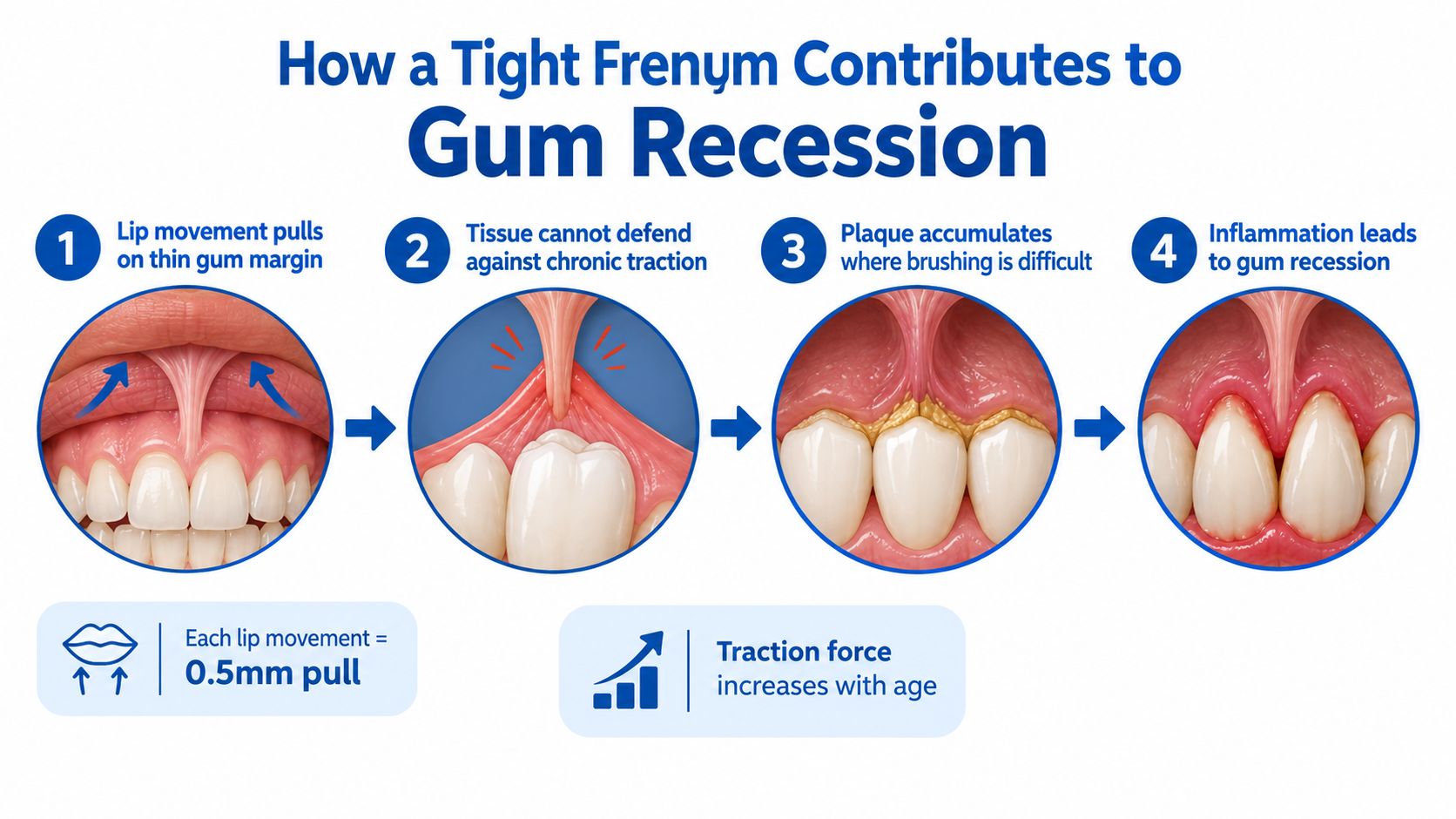
The possible mechanism is easier to understand as a chain of events. A frenum attached close to the gum edge can pull the tissue whenever the lip moves. If the gum is already thin or inflamed, repeated traction may make plaque control more difficult and may add stress to the area.
The possible chain of strain
- Lip movement creates tension. Smiling, speaking, eating, and lifting the lip can move the frenum.
- The gum margin receives repeated traction. A thin or poorly supported margin may have less resistance to that movement.
- Cleaning becomes harder. The band may pull the lip back toward a sensitive spot or create a shallow area where plaque remains.
- Inflammation develops. Plaque and calculus can irritate the gum and weaken its condition.
- Recession may progress. The margin can move toward the root, exposing more of the tooth.
The tent analogy helps explain why the process can be gradual. A stake pulling against the edge of a tent may not tear the fabric at once. Constant tension, poor weather, and an existing weak point make damage more likely. In the mouth, the comparable weak points may include thin gum tissue, inflammation, a tooth positioned outside the bony housing, or traumatic brushing.
Why the evidence needs a careful reading
The connection between a high or tight frenum and recession isn't certain in every patient. One review describes the frenum and recession relationship as inconclusive and controversial, while a newer review associates recession with several factors, including a high frenum, periodontal disease, smoking, alcohol use, and occlusal trauma, as discussed in this review of the frenum and recession relationship.
Gum recession can also involve:
- Plaque-induced inflammation and calculus
- Traumatic or overly forceful brushing
- Tooth malposition
- Restorative factors
- Uncontrolled orthodontic movement
- Periodontal breakdown from other causes
A review of recession causes lists these factors alongside high frenum attachment, which is why the multifactorial causes of gingival recession must be considered before surgery is selected.
The practical conclusion is straightforward. A frenum may be one suspect, but it shouldn't be treated as the entire case. A release is most logical when examination shows that the frenum is adding meaningful tension or interfering with hygiene, and when other causes are also being addressed.
When a Frenectomy Is Actually Indicated
A frenectomy is considered when the frenum's position or tension is linked to a specific periodontal or functional problem. The decision depends less on how dramatic the tissue looks and more on what the tissue is doing.
Situations that may support treatment
A periodontist may discuss frenectomy when an examination shows:
- Localized recession with active frenum pull: The gum moves noticeably when the lip is stretched, and the recession is concentrated around that attachment.
- A midline diastema: The frenum is associated with a persistent gap or tension between the front teeth.
- A flattened papilla and hygiene difficulty: The attachment interferes with brushing or allows plaque to remain near the gum edge.
- Inadequate attached gum: The area has limited firm gum and a shallow vestibule, leaving less tissue to resist movement.
- Persistent inflammation or pocketing: Food and plaque collect around the attachment despite appropriate cleaning.
Independent guidance from the American Academy of Pediatric Dentistry explains that a mandibular labial frenum can contribute to pocket formation, food and plaque accumulation, inflammation, recession, possible bone loss, and even tooth loss. The policy also states that controlling plaque-induced inflammation may reduce recession and the need for treatment, with surgery generally reserved for cases that continue despite oral hygiene management, according to this AAPD guidance on frenulum management.
Situations where other care may come first
| Clinical Situation | Typical First Approach |
|---|---|
| Recession with heavy plaque or active inflammation | Professional periodontal cleaning, home-care instruction, and inflammation control |
| Recession linked mainly to traumatic brushing | Modify brushing pressure, technique, and brush selection |
| A visible frenum without recession, tension, or hygiene problems | Observation and routine periodontal monitoring |
| Recession caused by tooth position or orthodontic movement | Evaluate tooth position and coordinate the broader treatment plan |
| Recession that continues after contributing factors are controlled and frenum pull remains | Consider frenectomy, sometimes with soft-tissue grafting |
Age also affects the context. A major review citing United States survey data reported recession at one or more sites in 37.8% of adults aged 30 to 39, rising to 90.4% of adults aged 80 to 90. The same review reported 88% prevalence among adults aged 65 and older and 50% among adults aged 18 to 64, as documented in this review of age-related gingival recession.
Those figures don't prove that a frenum caused a particular patient's recession. They show why recession is common among adults in New Castle and Kent Counties and why an evaluation must identify the specific driver. Treatment options and evaluations are available through the periodontal services offered in Wilmington, Newark, and Dover at Periodontal Associates of Delaware's periodontal services.
The Frenectomy Procedure at a Periodontal Practice
The procedure begins with diagnosis, not with cutting. A board-certified periodontist evaluates the frenum's attachment, the amount of attached gum, the recession pattern, pocket measurements, plaque and inflammation, tooth position, and the condition of the supporting bone.
What the appointment may involve
- Clinical examination: The clinician lifts or stretches the lip to observe whether the frenum moves the gum margin. Photographs, periodontal measurements, and imaging may help clarify the anatomy.
- Local anesthetic: The area is numbed before treatment. Patients may feel pressure or movement even when sharp sensation is reduced.
- Release or repositioning: The periodontist releases or reshapes the fibers so the lip or tongue no longer places the same tension on the gum.
- Site protection: Depending on the technique and anatomy, the area may receive sutures, a protective dressing, or specific home-care instructions.
- Follow-up: The clinician checks healing, hygiene, tissue stability, and the need for additional periodontal treatment.
A traditional frenectomy uses surgical instruments to remove or reposition the frenum. A laser-assisted approach may be appropriate in selected cases. Laser treatment can limit the surgical field and may support a minimally invasive plan, but the technique still has to match the anatomy and the clinical objective. A laser isn't a substitute for diagnosis.

How frenectomy fits into broader periodontal care
A patient with active gum disease may need scaling and root planing before a frenectomy. In selected cases, LANAP laser gum surgery using the FDA-cleared PerioLase MVP-7 may be part of periodontal disease treatment. Releasing a frenum while inflammation remains uncontrolled may leave a major cause of tissue damage untreated.
A patient with established root exposure may need a soft-tissue graft at the same time or during a later phase. Frenectomy can reduce the pull that threatens the area, while grafting may add tissue and provide root coverage. Neither procedure automatically replaces the other.
Patients from Wilmington, Newark, Pike Creek, Hockessin, Bear, Middletown, Dover, and surrounding communities can be evaluated at all three offices. For patients comparing treatment approaches, a discussion of laser gum treatment considerations can help clarify why technique, diagnosis, and the full periodontal condition matter more than the name of one procedure.
Advanced imaging is used when it answers a clinical question. i-CAT 3D cone beam imaging can help evaluate three-dimensional anatomy, while X-Nav dynamic surgical navigation is used for guided implant procedures when implants are part of a larger plan. These tools aren't required for every frenectomy, but they may be relevant when recession occurs alongside bone loss or missing teeth.
What to Expect After a Frenectomy and What It Can Achieve
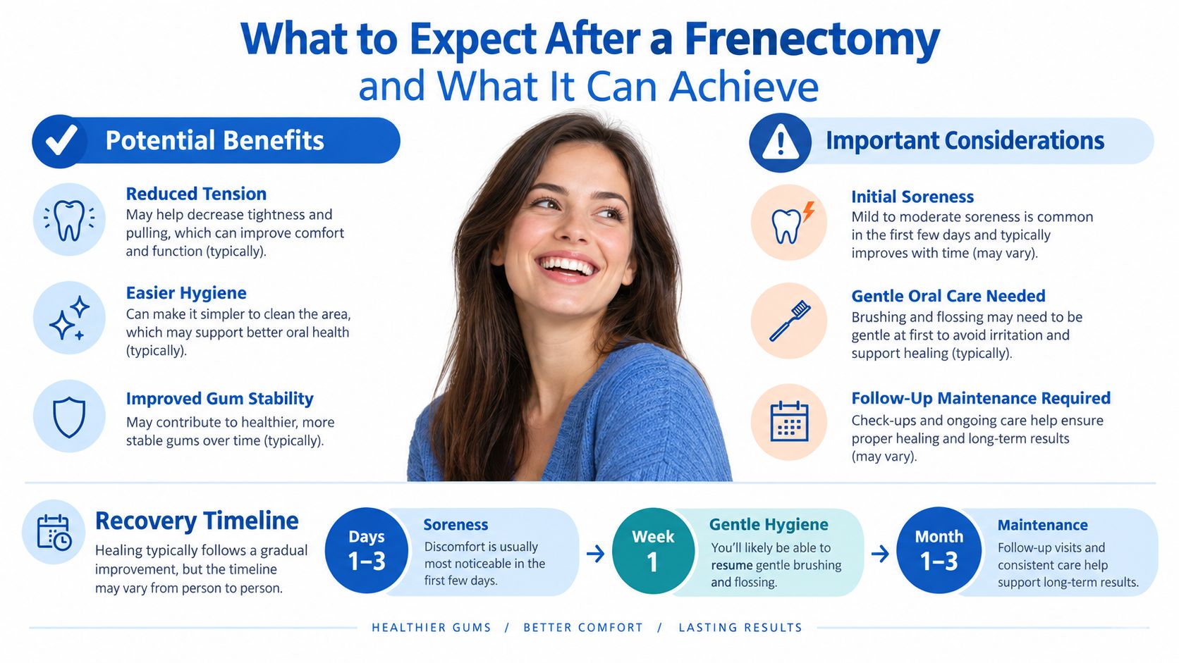
Many patients experience some soreness after a frenectomy, especially during the first part of healing. The treated area may feel tight or tender, and oral hygiene may need to be gentler around the site. The periodontist provides instructions for cleaning, eating, medication, and follow-up based on the procedure and the patient's medical history.
A typical recovery plan may include:
- Early healing: The area may be sore, and the lip or tongue should be moved according to the clinician's instructions.
- Gentle cleaning: Patients may need to avoid scrubbing directly over the healing site while still keeping nearby teeth clean.
- Periodontal maintenance: Follow-up visits help assess tissue stability, plaque control, inflammation, and any remaining recession.
- Additional treatment when needed: Root coverage or broader periodontal therapy may be recommended if tissue has already been lost.
What research suggests
A randomized clinical study involving patients with frenum-induced diastemas reported statistically significant improvements in gingival attachment, pocket depth, gingival recession, plaque index, and bleeding on probing after one year, as reported in this clinical study of periodontal changes after frenectomy. The findings support the idea that removing an abnormal frenum can improve soft-tissue health when the frenum is part of the disease mechanism.
The findings don't mean every patient will experience the same result. A separate clinical case report found that 12% of frenums recurred after surgery, which shows that anatomy, technique, healing, and ongoing forces can affect durability, as described in this report on frenectomy complications and recurrence.
The limit patients most need to understand
A frenectomy may stop or reduce a continuing pull, but it doesn't automatically replace gum that has already receded. If a root remains exposed, sensitivity or root coverage concerns may continue after the frenum is released. A graft may be needed when the treatment goal includes rebuilding the gum margin rather than just removing tension.
The most accurate takeaway is that frenectomy is often a first step in controlling a cause, not a guaranteed complete repair. Individual results vary, and a periodontist should evaluate the recession, frenum, inflammation, brushing habits, tooth position, and supporting tissues before a treatment decision is made.
Your Next Step Toward Healthier Gums
An aberrant frenum can contribute to localized recession, but recession is usually multifactorial. A periodontal evaluation can show whether the next step should be inflammation control, frenectomy, soft-tissue grafting, LANAP treatment, observation, or a combination of care.
Patients who feel anxious about gum surgery don't need to decide based on a photograph or a search result. A careful examination explains whether the tissue is pulling on the gum, whether the recession is progressing, and whether treatment can be limited to the smallest appropriate area. The practice's offices in Wilmington, Newark, and Dover use minimally invasive approaches when appropriate, along with tools such as the FDA-cleared PerioLase MVP-7 for LANAP and advanced imaging when the clinical situation calls for it.
Individual results vary. Each patient should consult a periodontist for advice based on the specific gum measurements, tissue thickness, medical history, and treatment goals.
Periodontal Associates of Delaware provides periodontal evaluations, frenectomy treatment, soft-tissue grafting, gum disease care, and related procedures for patients with recession or gum tension. Patients can visit Periodontal Associates of Delaware to request an appointment at the nearest Wilmington, Newark, or Dover office.